. 2003 May 15;22(10):2526-35.
doi: 10.1093/emboj/cdg238.

Xenopus Cut5 is essential for a CDK-dependent process in the initiation of DNA replication

Affiliations

Xenopus Cut5 is essential for a CDK-dependent process in the initiation of DNA replication

Yoshitami Hashimoto et al. EMBO J. .

Abstract

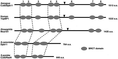
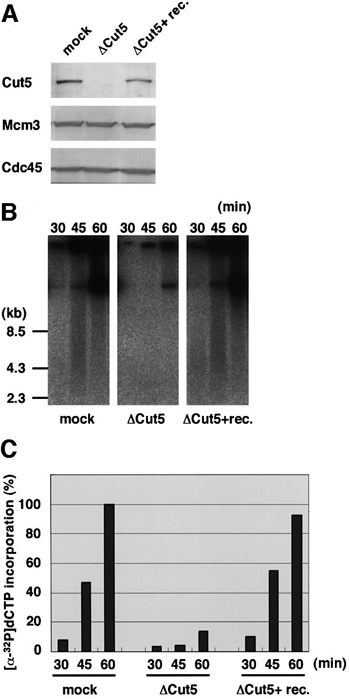
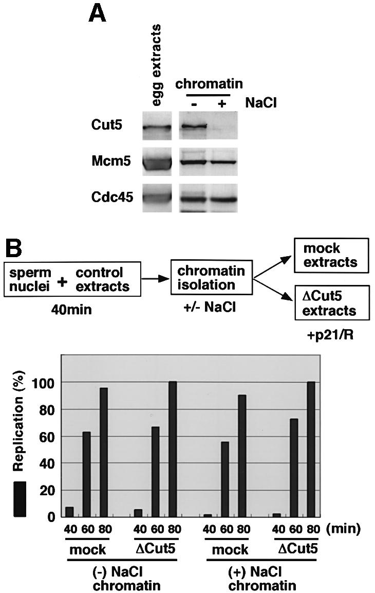
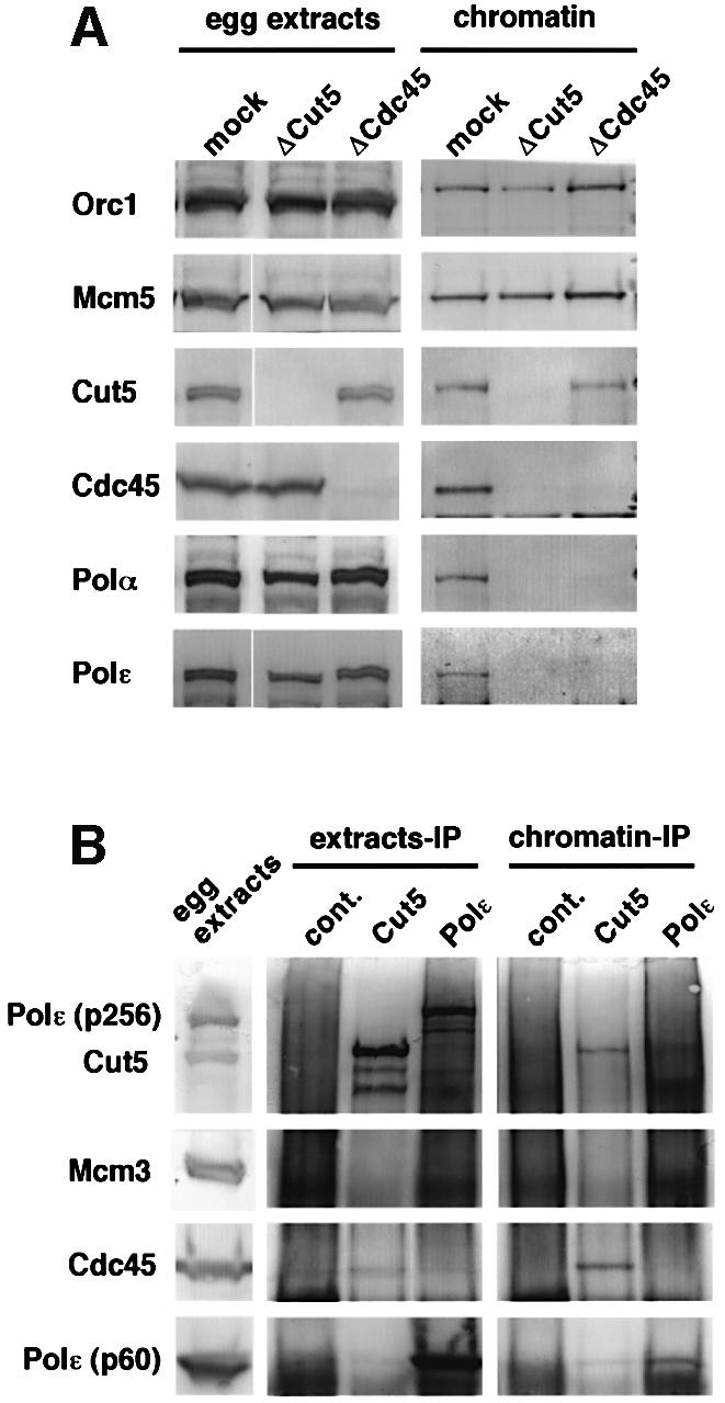
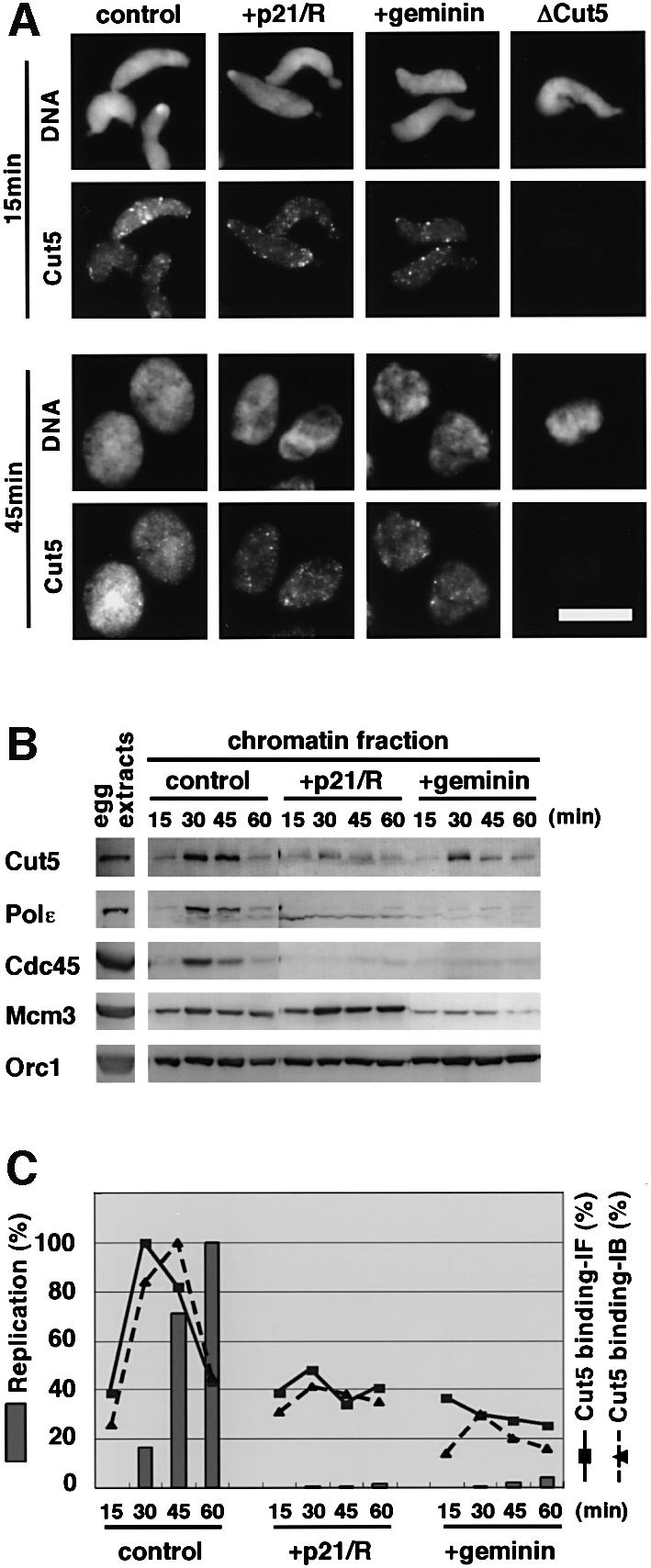
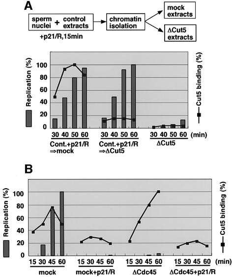
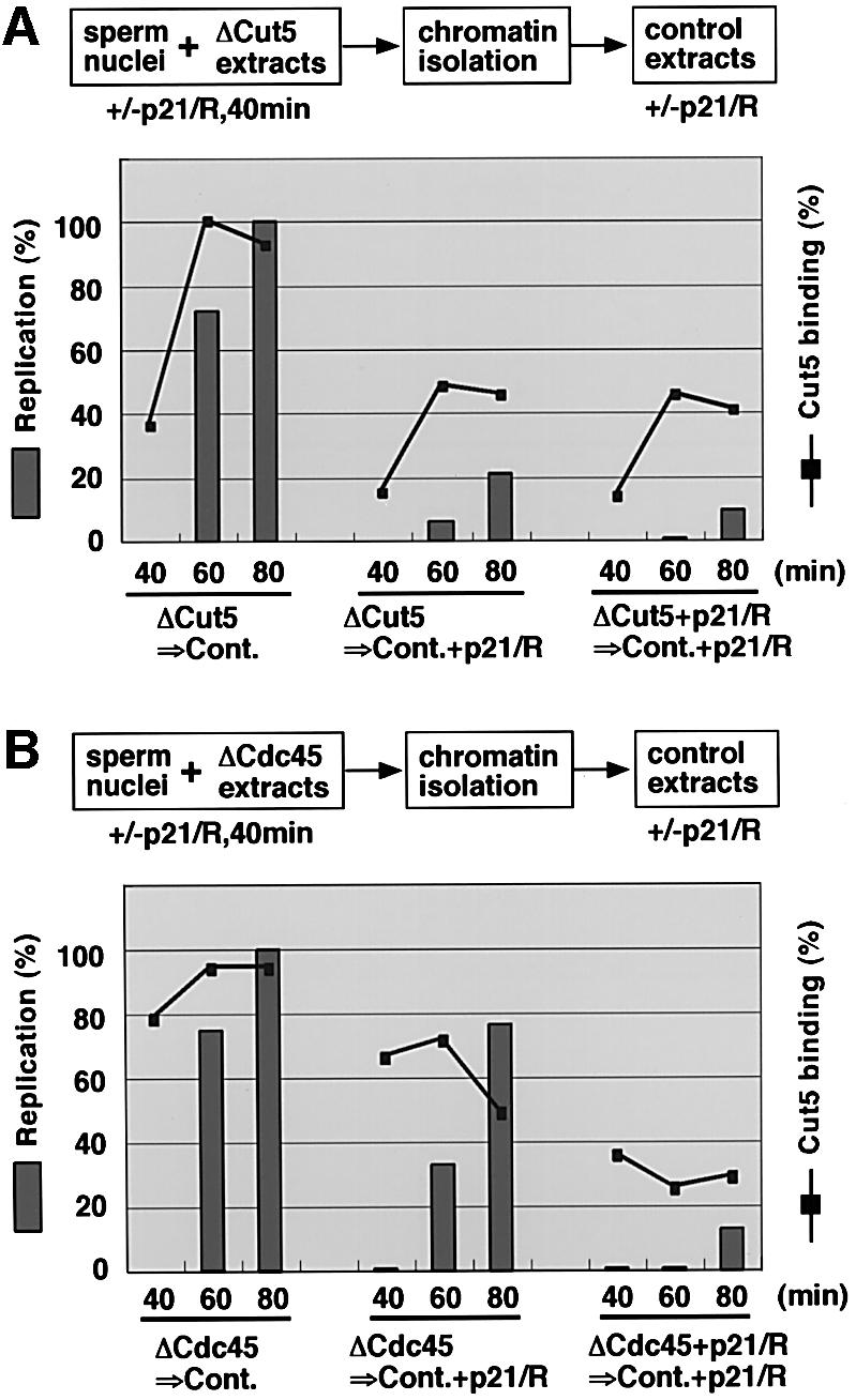
Fission yeast Cut5/Rad4 and its budding yeast homolog Dpb11 are required for both DNA replication and the S-phase checkpoint. Here, we have investigated the role of the Xenopus homolog of Cut5 in the initiation of DNA replication using Xenopus egg extracts. Xenopus Cut5, which shows sequence similarity to DmMus101 and HsTopBP1, is essential for DNA replication in the egg extracts. It is required for the chromatin binding of Cdc45 and DNA polymerases, but not for the formation of pre-replicative complexes or the elongation stage of DNA replication. The chromatin binding of Cut5 consists of two distinct modes. S-phase cyclin-dependent kinase (S-CDK)-independent binding is sufficient for DNA replication while S-CDK-dependent binding is dispensable. Further, S-CDK acts after the chromatin binding of Cut5 and before the binding of Cdc45. These results demonstrate that the chromatin binding of Cut5 is required for the action of S-CDK, which in turn triggers the formation of pre-initiation complexes of DNA replication.


Figures

None
Fig. 1. Schematic comparison of Cut5/Dpb11 homologs. Elliptical symbols represent BRCT domains. Dotted lines indicate highly conserved BRCT domains from yeast to human. BRCT domains 1–2 of the Xenopus protein share sequence similarity with the homologs in human (98%), Drosophila (54%), S.cerevisiae (30%) and S.pombe (47%), and BRCT domains 4–5 of the Xenopus protein share similarity with BRCT domains 4–5 of human (91%) and Drosophila (31%), and with BRCT domains 3–4 of S.cerevisiae (34%) and S.pombe (37%). Arrowheads indicate putative target sites of CDK conserved among Xenopus, human and Drosophila. Total numbers of amino acid residues are indicated at the right side.
None
Fig. 2. Time-courses of the chromatin binding of Cut5 and other replication proteins. (A) Specificity of anti-Cut5 antibody. S-phase egg extracts (1.5 µl) and 125 ng of recombinant Xenopus Cut5 protein (rec.Cut5) were resolved with SDS–PAGE and immunoblotted with anti-Xenopus Cut5 antibody. (B) Sperm nuclei were incubated in 50 µl of S-phase egg extract for the times indicated at 23°C. Chromatin fractions were isolated by centrifugation through a 10% sucrose layer and the isolated chromatin fractions and 1.5 µl of the extracts were resolved by SDS–PAGE, then immunoblotted with antibodies against the various replication proteins indicated.
None
Fig. 3. Requirement of Cut5 for DNA replication. (A) Immunodepletion of Cut5 from S-phase egg extracts. The egg extracts were treated with control and anti-Cut5 antibodies bound to a protein A matrix. Aliquots of mock-depleted (mock) and Cut5-depleted extracts with (ΔCut5+ rec.Cut5) or without (ΔCut5) 5 ng/µl of recombinant Cut5 were resolved by SDS–PAGE, then immunoblotted with antibodies against the proteins indicated in the figure. (B) Replication activity of Cut5- depleted extracts. Sperm nuclei were incubated in the treated egg extracts for the indicated times at 23°C. Replication activity was monitored as the incorporation of [α-32P]dCTP into sperm DNA. Replication products were subjected to agarose gel electrophoresis followed by autoradiography. (C) Relative intensities of autoradiography were analyzed and plotted against time, taking the highest value of the mock-depleted extracts as 100%.
None
Fig. 4. Requirement of Cut5 for elongation stages of DNA replication. (A) Selective removal of Cut5 from chromatin. Sperm nuclei were incubated in 50 µl of control egg extracts for 40 min at 23°C. The egg extracts (1.5 µl) and the chromatin fractions, isolated and washed with EB containing 0.0025% NP-40 in the absence (–) or presence (+) of 0.1 M NaCl, were subjected to immunoblotting. (B) Elongation activity of the isolated replicating chromatin fractions. The chromatin fractions prepared as in (A) were resuspended and incubated in mock- and Cut5-depleted egg extracts in the presence of 50 µg/ml GST–p21 and 100 µM roscovitine (+p21/R). Elongation activity was detected as the incorporation of Cy3-dCTP into chromatin DNA. The fluorescence intensities of 40 nuclei were analyzed. Relative average intensities per nucleus were then plotted against time, taking the maximal value as 100%.
None
Fig. 5. Requirement of Cut5 for the loading of Cdc45 onto chromatin. (A) Sperm nuclei were incubated in mock-, Cut5- and Cdc45-depleted egg extracts for 40 min at 23°C. For each extract, 1.5 µl depleted extract and the isolated chromatin fractions (chromatin) from 50 µl extracts were resolved with SDS–PAGE and immunoblotted. (B) Co-immunoprecipitation of Cdc45 with Cut5 on replicating chromatin. Sperm nuclei were incubated in S-phase egg extract in the presence of 50 µg/ml aphidicolin for 50 min at 23°C. Chromatin DNA was fragmented by sonication and the fragmented chromatin fractions were clarified by centrifugation. The egg extracts and the fragmented chromatin fractions (chromatin-IP) were immunoprecipitated with antibodies as indicated, then the immunoprecipitates were resolved with SDS–PAGE and immunoblotted.
None
Fig. 6. CDK-dependent and -independent binding of Cut5 to chromatin. (A) Immunofluorescent detection of Cut5 binding. Sperm nuclei were incubated in S-phase egg extracts in the absence (control) or in the presence of 50 µg/ml GST–p21 and 100 µM roscovitine (+p21/rosc.) or 15 µg/ml GST–geminin (+geminin) for the times indicated at 23°C. Cut5-depleted egg extracts (ΔCut5) were used as a negative control of immunofluorescence. Samples were treated with 0.25% NP-40 and then fixed with 3.7% formaldehyde. Nuclear localization of Cut5 was visualized with rabbit anti-Cut5 antibody followed by Alexa 488-labelled anti-rabbit IgG. DNA was visualized with Hoechst 33258. Scale bar 10 µm. (B) Time-course of chromatin binding of various replication proteins. Sperm nuclei were incubated in 50 µl of the extracts for the times indicated in the absence (control) or presence of p21 and roscovitine (+p21/rosc.) or 15 µg/ml GST–geminin (+geminin). One microliter of egg extract and the isolated chromatin fractions were resolved by SDS–PAGE, then immunoblotted with antibodies against various replication proteins. (C) Time-courses of DNA replication and chromatin binding of Cut5. Fluorescence images captured as in (A) and the immunoblotted bands in (B) were quantified in order to estimate the amounts of chromatin-bound Cut5. Replication activity was detected as the incorporation of Cy3-dCTP into DNA. The fluorescence intensities of 40 nuclei were analyzed using NIH Image software. Relative average intensities per nucleus were then plotted against time, taking the maximal value as 100% (Replication and Cut5 binding-IF). For immunoblotting, the intensities of immunoblotted bands of Cut5 were normalized against those of Orc1, a loading control for chromatin fractions, and ratios of the amounts of Cut5 to Orc1 were then plotted against time, taking the maximal value as 100% (Cut5 binding-IB).
None
Fig. 7. Requirement of chromatin binding of Cut5 for DNA replication. (A) Sperm nuclei were pre-incubated in S-phase egg extract in the presence of 50 µg/ml p21 and 100 µM roscovitine (p21/R) for 15 min at 23°C. The chromatin fractions were then isolated and incubated in mock- (Cont.+p21/R→mock) and Cut5-depleted (Cont.+p21/R→ ΔCut5) egg extracts at 23°C. At the times indicated, which include 15 min pre-incubation, samples were treated with NP-40, fixed with formaldehyde, and the amount of Cut5 bound to chromatin and the incorporation of Cy3-dCTP were analyzed as described in the legend for Figure 6. As a negative control, sperm nuclei were incubated in the Cut5-depleted egg extract throughout the time-course (ΔCut5). (B) S-CDK-dependent binding of Cut5 to chromatin in the absence of Cdc45. Sperm nuclei were incubated in mock- and Cdc45-depleted egg extracts with or without p21/roscovitine at 23°C. After incubation for the times indicated, samples were analyzed as in (A).
None
Fig. 8. Requirement of Cut5 and Cdc45 for the action of S-CDK. (A) Sperm nuclei were pre-incubated in Cut5-depleted egg extracts in the presence or absence of 50 µg/ml p21 and 100 µM roscovitine (p21/R) for 40 min at 23°C. The chromatin fractions were isolated and incubated in egg extracts in the absence (Cont.) or presence of p21/ roscovitine (Cont.+p21/R) at 23°C. After incubation for the times indicated, which include 40 min pre-incubation time, samples were treated with NP-40 and fixed with formaldehyde. The amount of Cut5 bound to chromatin and the incorporation of Cy3-dCTP were analyzed as described in the legend for Figure 6. (B) Similar experiments to those in (A) were performed, using Cdc45-depleted extracts instead of Cut5-depleted extracts.

Similar articles

Cited by

References

    1. Aparicio O.M., Stout,A.M. and Bell,S.P. (1999) Differential assembly of Cdc45p and DNA polymerases at early and late origins of DNA replication. Proc. Natl Acad. Sci. USA, 96, 9130–9135. - PMC - PubMed
    1. Araki H., Leem,S.H., Phongdara,A. and Sugino,A. (1995) Dpb11, which interacts with DNA polymerase II (ε) in Saccharomyces cerevisiae, has a dual role in S-phase progression and at a cell cycle checkpoint. Proc. Natl Acad. Sci. USA, 92, 11791–11795. - PMC - PubMed
    1. Bell S.P. and Dutta,A. (2002) DNA replication in eukaryotic cells. Annu. Rev. Biochem., 71, 333–374. - PubMed
    1. Blow J.J. and Laskey,R.A. (1986) Initiation of DNA replication in nuclei and purified DNA by a cell-free extract of Xenopus eggs. Cell, 47, 577–587. - PubMed
    1. Blow J.J., Gillespie,P.J., Francis,D. and Jackson,D.A. (2001) Replication origins in Xenopus egg extract are 5–15 kilobases apart and are activated in clusters that fire at different times. J. Cell Biol., 152, 15–25. - PMC - PubMed

Publication types

MeSH terms