

Computational imaging in cell biology
- PMID: 12743101
- PMCID: PMC2172925
- DOI: 10.1083/jcb.200302097
Computational imaging in cell biology
Abstract
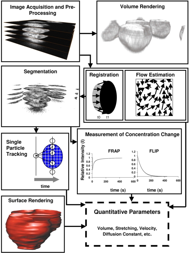
Microscopy of cells has changed dramatically since its early days in the mid-seventeenth century. Image analysis has concurrently evolved from measurements of hand drawings and still photographs to computational methods that (semi-) automatically quantify objects, distances, concentrations, and velocities of cells and subcellular structures. Today's imaging technologies generate a wealth of data that requires visualization and multi-dimensional and quantitative image analysis as prerequisites to turning qualitative data into quantitative values. Such quantitative data provide the basis for mathematical modeling of protein kinetics and biochemical signaling networks that, in turn, open the way toward a quantitative view of cell biology. Here, we will review technologies for analyzing and reconstructing dynamic structures and processes in the living cell. We will present live-cell studies that would have been impossible without computational imaging. These applications illustrate the potential of computational imaging to enhance our knowledge of the dynamics of cellular structures and processes.
Figures
References
-
- Adie, E.J., S. Kalinka, L. Smith, M.J. Francis, A. Marenghi, M.E. Cooper, M. Briggs, N.P. Michael, G. Milligan, and S. Game. 2002. A pH-sensitive fluor, CypHer 5, used to monitor agonist-induced G protein-coupled receptor internalization in live cells. Biotechniques. 33:1152–1154, 1156–1157. - PubMed
-
- Allen, R.D., and N.S. Allen. 1983. Video-enhanced microscopy with a computer frame memory. J. Microsc. 129:3–17. - PubMed
-
- Anandan, P. 1989. A computational framework and an algorithm for the measurement of visual motion. Int. J. Comp. Vis. 2:283–310.
-
- Arun, K.S., T.S. Huang, and S.D. Blostein. 1987. Least square fitting of two 3D point sets. Transactions of Pattern Analysis and Machine Intelligence. 9:698–700. - PubMed
Publication types
MeSH terms
LinkOut - more resources
Full Text Sources
Other Literature Sources
