Spindle orientation in Saccharomyces cerevisiae depends on the transport of microtubule ends along polarized actin cables
- PMID: 12743102
- PMCID: PMC2172944
- DOI: 10.1083/jcb.200302030
Spindle orientation in Saccharomyces cerevisiae depends on the transport of microtubule ends along polarized actin cables
Abstract
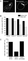
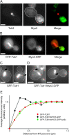
Microtubules and actin filaments interact and cooperate in many processes in eukaryotic cells, but the functional implications of such interactions are not well understood. In the yeast Saccharomyces cerevisiae, both cytoplasmic microtubules and actin filaments are needed for spindle orientation. In addition, this process requires the type V myosin protein Myo2, the microtubule end-binding protein Bim1, and Kar9. Here, we show that fusing Bim1 to the tail of the Myo2 is sufficient to orient spindles in the absence of Kar9, suggesting that the role of Kar9 is to link Myo2 to Bim1. In addition, we show that Myo2 localizes to the plus ends of cytoplasmic microtubules, and that the rate of movement of these cytoplasmic microtubules to the bud neck depends on the intrinsic velocity of Myo2 along actin filaments. These results support a model for spindle orientation in which a Myo2-Kar9-Bim1 complex transports microtubule ends along polarized actin cables. We also present data suggesting that a similar process plays a role in orienting cytoplasmic microtubules in mating yeast cells.
Figures


Similar articles
-
Gamma-tubulin is required for proper recruitment and assembly of Kar9-Bim1 complexes in budding yeast.Mol Biol Cell. 2006 Oct;17(10):4420-34. doi: 10.1091/mbc.e06-03-0245. Epub 2006 Aug 9. Mol Biol Cell. 2006. PMID: 16899509 Free PMC article.
-
The role of the proteins Kar9 and Myo2 in orienting the mitotic spindle of budding yeast.Curr Biol. 2000 Nov 30;10(23):1497-506. doi: 10.1016/s0960-9822(00)00837-x. Curr Biol. 2000. PMID: 11114516
-
Cdk1-Clb4 controls the interaction of astral microtubule plus ends with subdomains of the daughter cell cortex.Genes Dev. 2004 Jul 15;18(14):1709-24. doi: 10.1101/gad.298704. Genes Dev. 2004. PMID: 15256500 Free PMC article.
-
Kar9 asymmetrical loading on spindle poles mediates proper spindle alignment in budding yeast.Dev Cell. 2003 Mar;4(3):289-90. doi: 10.1016/s1534-5807(03)00065-0. Dev Cell. 2003. PMID: 12636909 Review.
-
Why do peroxisomes associate with the cytoskeleton?Biochim Biophys Acta. 2016 May;1863(5):1019-26. doi: 10.1016/j.bbamcr.2015.11.022. Epub 2015 Nov 23. Biochim Biophys Acta. 2016. PMID: 26616035 Review.
Cited by
-
Cargo Release from Myosin V Requires the Convergence of Parallel Pathways that Phosphorylate and Ubiquitylate the Cargo Adaptor.Curr Biol. 2020 Nov 16;30(22):4399-4412.e7. doi: 10.1016/j.cub.2020.08.062. Epub 2020 Sep 10. Curr Biol. 2020. PMID: 32916113 Free PMC article.
-
Yeast formin Bni1p has multiple localization regions that function in polarized growth and spindle orientation.Mol Biol Cell. 2012 Feb;23(3):412-22. doi: 10.1091/mbc.E11-07-0631. Epub 2011 Dec 7. Mol Biol Cell. 2012. PMID: 22160598 Free PMC article.
-
Finding the cell center by a balance of dynein and myosin pulling and microtubule pushing: a computational study.Mol Biol Cell. 2010 Dec;21(24):4418-27. doi: 10.1091/mbc.E10-07-0627. Epub 2010 Oct 27. Mol Biol Cell. 2010. PMID: 20980619 Free PMC article.
-
The cyclin-dependent kinase Cdc28p regulates multiple aspects of Kar9p function in yeast.Mol Biol Cell. 2007 Apr;18(4):1187-202. doi: 10.1091/mbc.e06-04-0360. Epub 2007 Jan 24. Mol Biol Cell. 2007. PMID: 17251549 Free PMC article.
-
Actomyosin-dependent cortical dynamics contributes to the prophase force-balance in the early Drosophila embryo.PLoS One. 2011 Mar 31;6(3):e18366. doi: 10.1371/journal.pone.0018366. PLoS One. 2011. PMID: 21483831 Free PMC article.
References
-
- Beach, D.L., J. Thibodeaux, P. Maddox, E. Yeh, and K. Bloom. 2000. The role of the proteins Kar9 and Myo2 in orienting the mitotic spindle of budding yeast. Curr. Biol. 10:1497–1506. - PubMed
-
- Bienz, M. 2001. Spindles cotton on to junctions, APC and EB1. Nat. Cell Biol. 3:E67–E68. - PubMed
-
- Bloom, K. 2000. It's a kar9ochore to capture microtubules. Nat. Cell Biol. 2:E96–E98. - PubMed
Publication types
MeSH terms
Substances
Grants and funding
LinkOut - more resources
Full Text Sources
Other Literature Sources
Molecular Biology Databases
Miscellaneous

