. 2003 May 12;161(3):489-95.
doi: 10.1083/jcb.200303138.

Neurofilament heavy chain side arm phosphorylation regulates axonal transport of neurofilaments

Affiliations

Neurofilament heavy chain side arm phosphorylation regulates axonal transport of neurofilaments

Steven Ackerley et al. J Cell Biol. .

Abstract

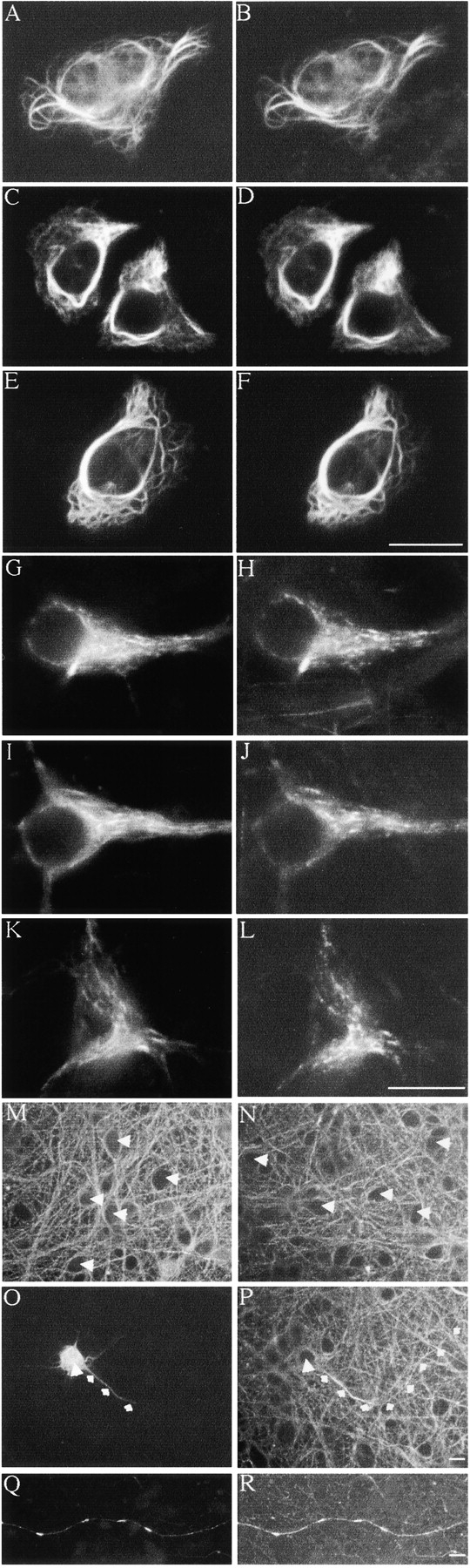
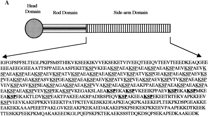
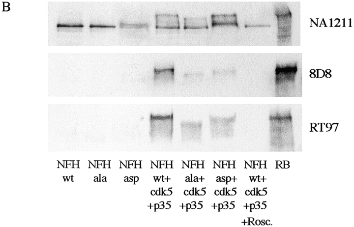
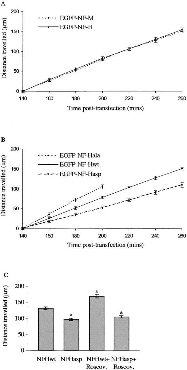
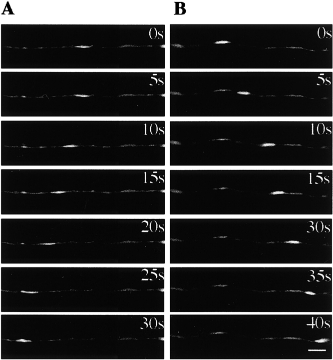
Neurofilaments possess side arms that comprise the carboxy-terminal domains of neurofilament middle and heavy chains (NFM and NFH); that of NFH is heavily phosphorylated in axons. Here, we demonstrate that phosphorylation of NFH side arms is a mechanism for regulating transport of neurofilaments through axons. Mutants in which known NFH phosphorylation sites were mutated to preclude phosphorylation or mimic permanent phosphorylation display altered rates of transport in a bulk transport assay. Similarly, application of roscovitine, an inhibitor of the NFH side arm kinase Cdk5/p35, accelerates neurofilament transport. Analyses of neurofilament movement in transfected living neurons demonstrated that a mutant mimicking permanent phosphorylation spent a higher proportion of time pausing than one that could not be phosphorylated. Thus, phosphorylation of NFH slows neurofilament transport, and this is due to increased pausing in neurofilament movement.


Figures

Figure 1.
Figure 1.
GFP–NFH assembly and phosphorylation mimics that of endogenous NFH. (A–F) SW13− cells transfected with NFL + NFM + NFH + either GFP–NFHwt (A and B), GFP–NFHala (C and D), or GFP–NFHasp (E and F). (G–L) Cortical neurons transfected with GFP–NFHwt (G and H), GFP–NFHala (I and J), or GFP–NFHasp (K and L). GFP–NFH was detected via the GFP tag in A, C, E, G, I, and K. NFL was detected using antibody NR4 in B, D, F, H, J, and L. A–L were visualized 16 h after transfection, but similar images of cortical neurons were obtained at earlier (140–260 min) and later (48 h) times. (M and N) Cortical neurons stained with antibodies 8D8 (M) and RT97 (N); unstained cell bodies are arrowed. (O–R) Cortical neurons transfected with GFP–NFHwt and costained with RT97. O and P are visualized 200 min after transfection, and Q and R are visualized 48 h after transfection. O and Q show GFP–NFHwt via the GFP tag; P and R show RT97 labeling. Large arrow shows cell body, and small arrows show trace axon of a transfected cell. Note the increased RT97 labeling in regions of axons where GFP–NFHwt is present, but also note the absence of RT97 labeling in cell bodies. Bars, 20 μm.
Figure 2.
Figure 2.
Structure and phosphorylation of rat NFH by Cdk5/p35. (A) Schematic of rat NFH with KSP sites underlined and mutated sites shown in bold. (B) Phosphorylation of NFHwt, NFHala, and NFHasp by Cdk5/p35 in transfected COS cells. Cells were transfected with NFHwt, NFHala, or NFHasp ± Cdk5/p35 as indicated, and the samples were probed on immunoblots with antibodies NA1211, 8D8, and RT97. RB is a sample of rat brain. 8D8 and RT97 both recognize phosphorylated NFH side arms; NA1211 is an NFH phosphorylation-independent antibody. Roscovitine (Rosc.) was applied 16 h before harvesting.
Figure 2.
Figure 2.
Structure and phosphorylation of rat NFH by Cdk5/p35. (A) Schematic of rat NFH with KSP sites underlined and mutated sites shown in bold. (B) Phosphorylation of NFHwt, NFHala, and NFHasp by Cdk5/p35 in transfected COS cells. Cells were transfected with NFHwt, NFHala, or NFHasp ± Cdk5/p35 as indicated, and the samples were probed on immunoblots with antibodies NA1211, 8D8, and RT97. RB is a sample of rat brain. 8D8 and RT97 both recognize phosphorylated NFH side arms; NA1211 is an NFH phosphorylation-independent antibody. Roscovitine (Rosc.) was applied 16 h before harvesting.
Figure 3.
Figure 3.
Analyses of GFP–NFH transport in transfected neurons. (A) Distances travelled by GFP–NFHwt and GFP–NFM at 140–260 min after transfection. One-way ANOVA tests showed no significant differences between GFP–NFHwt and GFP–NFM transport at any time point. (B) Distances travelled by GFP–NFHwt, GFP–NFHala, and GFP–NFHasp at 140–260 min after transfection. One-way ANOVA tests showed significant differences between GFP–NFHwt and GFP–NFHala (P = 0.045) and between GFP–NFHala and GFP–NFHasp (P = 0.001) at the 160-min time point. GFP–NFHwt and GFP–NFHasp displayed significant differences at the 180-min time point (P = 0.005). At later time points, significant differences between all three GFP–NFH proteins were observed (P < 0.001). (C) Histogram shows distance travelled by GFP–NFHwt and GFP–NFHasp 240 min after transfection in either 20 μM roscovitine (Roscov.) or vehicle-treated neurons. Roscovitine or vehicle was applied 130 min after transfection. An asterisk indicates treatments that display significant differences (P < 0.001) compared with vehicle-treated GFP–NFHwt, as analyzed by One-way ANOVA tests. No significant difference of GFP–NFHasp transport was observed between vehicle- and roscovitine-treated neurons. As detailed previously (Ackerley et al., 2000), the distances travelled by GFP–NFHwt/ala/asp at the first (140 min) time point are all adjusted to zero, so as to facilitate comparisons between experiments, and do not represent the actual distances travelled from cell bodies. Each data set shown for A–C is from one representative experiment, and error bars are the SEM.
Figure 4.
Figure 4.
GFP–NFH movement in living neurons. (A and B) Stills from movies of GFP–NFHwt movement in anterograde (A) and retrograde (B) directions. The directions of movement (anterograde, right to left; retrograde, left to right) are due to the orientation of axons when the images were captured. Camera exposure times in seconds are shown. Bar is 5 μm. (C) Representative movement characteristics of GFP–NFHwt, GFP–NFHala, and GFP–NFHasp as indicated. Each point represents the distance moved by the filament from its starting position measured along the axon in micrometers; exposures are every 5 s. Asterisks show pauses in movement of GFP–NFHasp.
Figure 4.
Figure 4.
GFP–NFH movement in living neurons. (A and B) Stills from movies of GFP–NFHwt movement in anterograde (A) and retrograde (B) directions. The directions of movement (anterograde, right to left; retrograde, left to right) are due to the orientation of axons when the images were captured. Camera exposure times in seconds are shown. Bar is 5 μm. (C) Representative movement characteristics of GFP–NFHwt, GFP–NFHala, and GFP–NFHasp as indicated. Each point represents the distance moved by the filament from its starting position measured along the axon in micrometers; exposures are every 5 s. Asterisks show pauses in movement of GFP–NFHasp.

References

    1. Ackerley, S., A.J. Grierson, J. Brownlees, P. Thornhill, B.H. Anderton, P.N. Leigh, C.E. Shaw, and C.C.J. Miller. 2000. Glutamate slows axonal transport of neurofilaments in transfected neurons. J. Cell Biol. 150:165–175. - PMC - PubMed
    1. Bajaj, N.P., and C.C.J. Miller. 1997. Phosphorylation of neurofilament heavy-chain side-arm fragments by cyclin-dependent kinase-5 and glycogen synthase kinase-3a in transfected cells. J. Neurochem. 69:737–743. - PubMed
    1. Brownlees, J., A. Yates, N.P. Bajaj, D. Davis, B.H. Anderton, P.N. Leigh, C.E. Shaw, and C.C.J. Miller. 2000. Phosphorylation of neurofilament heavy chain side-arms by stress activated protein kinase-1b/Jun N-terminal kinase-3. J. Cell Sci. 113:401–407. - PubMed
    1. Brownlees, J., S. Ackerley, A.J. Grierson, N.J. Jacobsen, K. Shea, B.H. Anderton, P.N. Leigh, C.E. Shaw, and C.C. Miller. 2002. Charcot-Marie-Tooth disease neurofilament mutations disrupt neurofilament assembly and axonal transport. Hum. Mol. Genet. 11:2837–2844. - PubMed
    1. Collard, J.-F., F. Cote, and J.-P. Julien. 1995. Defective axonal transport in a transgenic mouse model of amyotrophic lateral sclerosis. Nature. 375:61–64. - PubMed

Publication types

MeSH terms