. 2003 May 12;161(3):557-70.
doi: 10.1083/jcb.200211122.

Alpha-adducin dissociates from F-actin and spectrin during platelet activation

Affiliations

Alpha-adducin dissociates from F-actin and spectrin during platelet activation

Kurt L Barkalow et al. J Cell Biol. .

Abstract

Aspectrin-based skeleton uniformly underlies and supports the plasma membrane of the resting platelet, but remodels and centralizes in the activated platelet. alpha-Adducin, a phosphoprotein that forms a ternary complex with F-actin and spectrin, is dephosphorylated and mostly bound to spectrin in the membrane skeleton of the resting platelet at sites where actin filaments attach to the ends of spectrin molecules. Platelets activated through protease-activated receptor 1, FcgammaRIIA, or by treatment with PMA phosphorylate adducin at Ser726. Phosphoadducin releases from the membrane skeleton concomitant with its dissociation from spectrin and actin. Inhibition of PKC blunts adducin phosphorylation and release from spectrin and actin, preventing the centralization of spectrin that normally follows cell activation. We conclude that adducin targets actin filament ends to spectrin to complete the assembly of the resting membrane skeleton. Dissociation of phosphoadducin releases spectrin from actin, facilitating centralization of spectrin, and leads to the exposure of barbed actin filament ends that may then participate in converting the resting platelet's disc shape into its active form.


Figures
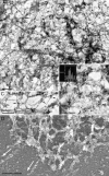
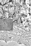
Figure 1.
Figure 1.
α-Adducin localizes to periodic sites that are likely to be the ends of spectrin tetramers. (A) Resting platelet cytoskeletons were labeled with 10 nm gold coated with goat anti–rabbit IgG. Gold is found in small clusters separated by ∼200 nm at the ends of triangular pores (B) that are evenly distributed across the membrane skeleton. The distance between adjacent clusters is 201 ± 46 nm (n = 55). The inset shows the frequency length distribution of the intercluster distance. (C) A more open area of the membrane skeleton showing that anti-adducin labeling is found at the intersection of fibers. (D) Resting platelet cytoskeletons were sedimented onto glass coverslips without fixation to fracture the cytoskeleton and thus separate the spectrin-rich membrane skeleton from the core of F-actin. This image shows the membrane skeleton consisting primarily of spectrin filaments. Spectrin filaments are distinguished from actin, which has a rope-like appearance here, by virtue of labeling with myosin S1. Adducin is found almost exclusively in the spectrin-rich region. Arrows indicate the polarity of F-actin by pointing in the direction of the slow-growing or “pointed” end. Bars, 200 nm.
Figure 2.
Figure 2.
Localization of spectrin in the cytoskeleton of a spread platelet by immunoelectron microscopy. (A and B) high magnification images of those indicated in the inset. Dense binding of 10 nm immunogold anti-spectrin is found in the center of the cytoskeleton (A), whereas only sparse gold labeling is found in the rest of the cytoskeleton (B). (C and D) Immunogold labeling of platelet cytoskeletons shows little adducin remaining in the center (C) or in the cortex (D) after activation. Bars, 200 nm.
Figure 2.
Figure 2.
Localization of spectrin in the cytoskeleton of a spread platelet by immunoelectron microscopy. (A and B) high magnification images of those indicated in the inset. Dense binding of 10 nm immunogold anti-spectrin is found in the center of the cytoskeleton (A), whereas only sparse gold labeling is found in the rest of the cytoskeleton (B). (C and D) Immunogold labeling of platelet cytoskeletons shows little adducin remaining in the center (C) or in the cortex (D) after activation. Bars, 200 nm.
Figure 3.
Figure 3.
Immunofluorescent localization of adducin, spectrin, and F-actin in platelets spread on glass. The protein stained and fluorophore of the secondary antibody are indicated in each panel. Samples pretreated with 5 μM GF or 2 μM cytochalasin B are indicated. “Cytoskeleton” denotes platelets first extracted with Triton X-100 before fixation. All other panels show intact and fixed platelets. Bar, 5 μm.
Figure 4.
Figure 4.
Immunofluorescent localization of adducin, spectrin, and F-actin as platelets spread for 1.5 or 5 min. The protein stained and fluorophore of the secondary antibody is indicated in each panel. Samples treated with GF are indicated. Panels showing resting platelets are located in the top left. Bar, 5 μm. GF treatment reduces the separation of actin from spectrin and adducin. The two graphs in the bottom left quantify the amount of colocalization of F-actin staining with spectrin (top graph) or adducin (bottom graph) staining during normal platelet spreading (squares with black lines) or spreading in the presence of 5 μM GF109203X (diamonds with red lines) with respect to the incubation time (mean ± SD, n = 10).
Figure 5.
Figure 5.
α-Adducin is bound to the actin cytoskeletons of resting platelets and is phosphorylated and released from the cytoskeletons of active platelets. (A–C) Phosphorylation of adducin at Ser726 follows platelet activation by PAR-1 (A), FcγRIIA (B), and PMA (C). The kinetics of adducin phosphorylation are plotted (mean ± SD, n = 3). Insets (A and C) show representative Ser726 phosphoimmunoblots ± GF for PAR-1 and PMA activation. (D–F) The amount of adducin associated with F-actin in 215,000 g pellets of permeabilized platelets (squares) activated by PAR-1 (D), FcγRIIA (E), or PMA (F) is compared with platelets treated before permeabilization with either GF (diamonds, D–F) or EGTA-AM (circles, D and F).
Figure 6.
Figure 6.
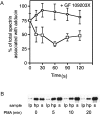
α-Adducin associates with spectrin in resting platelets, but dissociates during platelet activation. (A) Percentage of total spectrin that immunoprecipitates with adducin in resting platelets or platelets activated with 25 μM TRAP (squares). Less spectrin associates with α-adducin after platelet activation. Dissociation of spectrin from adducin is prevented when platelets are incubated with the PKC inhibitor GF 109203X (diamonds). (B) Distribution of adducin in the cytoskeleton (lp, 15,000 g low speed pellet), membrane skeleton (hp, 215,000 g high speed pellet), and soluble (s, high speed supernatant) phase of PMA-stimulated platelets permeabilized with Triton X-100 and detected by immunoblot.
Figure 7.
Figure 7.
Treatment of permeabilized, washed platelets with the catalytic subunit of PKC or PI3,4P2 micelles releases α-adducin from the cytoskeleton. (A) Platelets were extracted with 0.4% OG and washed twice in PHEM without OG. They were treated with the catalytic subunit of PKC (Sigma-Aldrich) at the indicated times, and then centrifuged to generate pellet (p) and supernatant (s) fractions. Immunoblots of total cytoskeletal α-adducin (top immunoblot) or Ser726 phospho–α-adducin (bottom immunoblot) are shown. (B) Immunoblots of total α-adducin in cytoskeletal pellets (p) and soluble fractions (s) of permeabilized platelets treated with phosphatidylserine (PS), PI3,4P2, or PI4,5P2.
Figure 8.
Figure 8.
Actin assembly and barbed-end exposure are diminished by inhibiting PKC. (A and B) Cytoskeletal changes are temporally linked to the phosphorylation and release of adducin from the cytoskeleton. (A) F-actin content and (B) barbed-end exposure in resting platelets and platelets stimulated through PAR-1 in the absence (squares) or the presence (diamonds) of GF. (C and D) Comparison of F-actin content (C) and free barbed ends (D) in platelets activated through FcγRIIA in the absence (squares) or presence (diamonds) of GF. Both are reduced by 50–70% in platelets stimulated through FcγRIIA when PKC is inhibited. (E and F) Platelets stimulated by 100 nM PMA exhibit marked inhibition of F-actin assembly (E) and barbed-end exposure (F) when pretreated with GF (diamonds), compared with controls (squares).
Figure 9.
Figure 9.
Inhibition of PKC by GF does not alter the activation of gelsolin, capZ, or Arp2/3 in PAR-1–ligated platelets. Results from platelets stimulated by PAR-1 ligation or addition of PMA are shown. PMA does not induce an association of gelsolin or Arp2/3 with the cytoskeleton, but capZ does associate with the cytoskeleton in a PKC-dependent fashion. All data are expressed as a percentage of maximal association with the cytoskeleton (mean ± SD, n = 3).
Figure 10.
Figure 10.
Diagram summarizing the reorganization of F-actin, spectrin, and adducin during platelet spreading. (Left) Normal spreading. The resting platelet has a spectrin lattice that laminates the cytoplasmic face of its plasma membrane. Adducin molecules sit at the intersections of spectrin tetramers and bind F-actin barbed ends. Changes in the adducin–actin–spectrin interaction occur as platelets become active. Platelet activation (early events) results in gelsolin-mediated F-actin severing and adducin release from spectrin–actin, which destabilizes the membrane skeleton. (Later events) Actin polymerization and centralization of spectrin–dephosphoadducin–F-actin follow the fragmentation phase of platelet activation. (Right) PKC inhibition slows spectrin centralization. F-actin is severed by gelsolin, but adducin is not phosphorylated and cannot release from spectrin–actin. Actin nucleation sites remain associated with the membrane skeleton because F-actin fragments remain bound to spectrin. This enhances actin assembly in the region of the membrane skeleton, which does not condense and remains dispersed in the center of the activated platelet. Spectrin, actin, and adducin are defined in the diagram.

Similar articles

Cited by

References

    1. Amano, M., A. Chihara, K. Kimura, Y. Fukata, N. Nakamura, Y. Matsuura, and K. Kaibuchi. 1997. Formation of actin stress fibers and focal adhesions enhanced by rho-kinase. Science. 275:1308–1311. - PubMed
    1. Barkalow, K., W. Witke, D. Kwiatkowski, and J. Hartwig. 1996. Coordinated regulation of platelet actin filament barbed ends by gelsolin and capping protein. J. Cell Biol. 134:389–399. - PMC - PubMed
    1. Bennett, V., K. Gardner, and J.P. Steiner. 1988. Brain adducin: a protein kinase C substrate that may mediate site-directed assembly at the spectrin-actin junction. J. Biol. Chem. 263:5860–5869. - PubMed
    1. Boyles, J., J.E.B. Fox, D.R. Phillips, and P.E. Stenberg. 1985. Organization of the cytoskeleton in resting, discoid platelets: preservation of actin filaments by a modified fixation that prevents osmium damage. J. Cell Biol. 101:1463–1472. - PMC - PubMed
    1. Falet, H., and F. Rendu. 1994. Calcium mobilisation controls tyrosine protein phosphorylation independently of the activation of protein kinase C in human platelets. FEBS Lett. 345:87–91. - PubMed

Publication types

MeSH terms