

Axonal transport and sorting of herpes simplex virus components in a mature mouse visual system
- PMID: 12743269
- PMCID: PMC155024
- DOI: 10.1128/jvi.77.11.6117-6126.2003
Axonal transport and sorting of herpes simplex virus components in a mature mouse visual system
Abstract
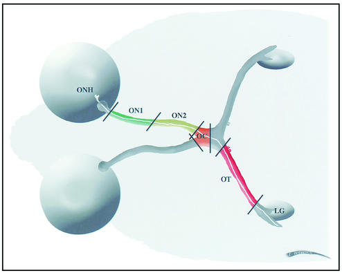
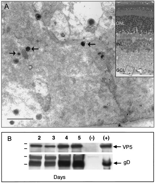
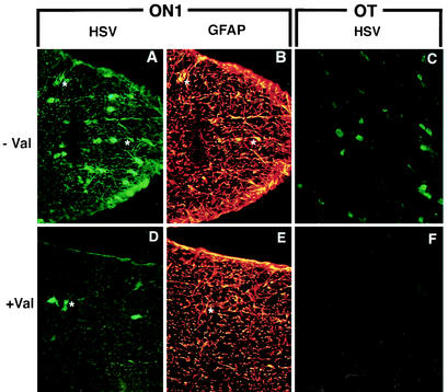
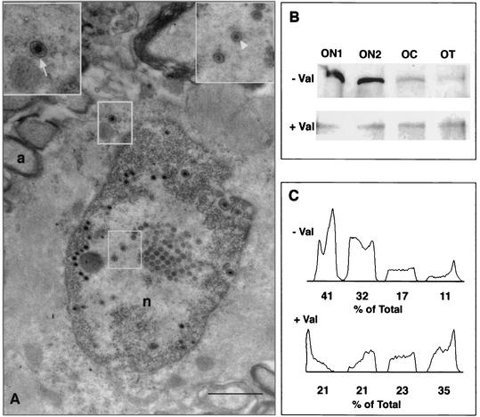
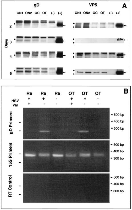
The time course for delivery and transport of two major proteins of herpes simplex virus (HSV) has been determined for mature mouse retinal ganglion cell axons in vivo. Twenty-four hours after intravitreal injection of HSV, valacyclovir was introduced into the drinking water of the mice to inhibit subsequent viral replication. Without treatment, viral spread and replication in periaxonal glial cells confound study of axonal transport. At 2 to 5 days after infection, the animals were sacrificed and contiguous segments of the optic pathway were removed. Immunofluorescence microscopy indicated that the number of infected astrocytes was reduced in the proximal optic nerve and eliminated in the optic tract. Western blots of the retina with antibodies for envelope and capsid components, glycoprotein D (gD) and VP5, respectively, revealed that both components were expressed in retinal homogenates by 2 days. Results of reverse transcription-PCR indicated that there was no gD mRNA present in the treated optic tract 5 days after infection. Therefore, we conclude that gD is transcribed from viral mRNA in the retinal ganglion cell bodies. The gD accumulated in the proximal ganglion cell axon by 2 days and reached the most distal segment after 3 days. The VP5 first appeared in the proximal axons at 4 days, about 48 h after the appearance of gD. Thus, gD entered the axon earlier and independent of VP5. These finding confirm the subassembly model of viral transport in neurons and suggest that there is a 4- to 5-day window for initiation of effective antiviral treatment with valacyclovir.
Figures
Similar articles
-
Herpes simplex virus type 1 glycoprotein e is required for axonal localization of capsid, tegument, and membrane glycoproteins.J Virol. 2005 Nov;79(21):13362-72. doi: 10.1128/JVI.79.21.13362-13372.2005. J Virol. 2005. PMID: 16227258 Free PMC article.
-
[Herpes simplex virus latency, reactivation, and a new antiviral therapy for herpetic keratitis].Nippon Ganka Gakkai Zasshi. 2008 Mar;112(3):247-64; discussion 265. Nippon Ganka Gakkai Zasshi. 2008. PMID: 18411713 Review. Japanese.
-
Valacyclovir for the prevention of recurrent herpes simplex virus eye disease after excimer laser photokeratectomy.Trans Am Ophthalmol Soc. 2000;98:285-303. Trans Am Ophthalmol Soc. 2000. PMID: 11190029 Free PMC article.
-
Temporal expression of herpes simplex virus type 1 mRNA in murine retina.Curr Eye Res. 2004 Aug-Sep;29(2-3):191-4. doi: 10.1080/02713680490504597. Curr Eye Res. 2004. PMID: 15512966
-
Herpes simplex virus keratitis: an update of the pathogenesis and current treatment with oral and topical antiviral agents.Clin Exp Ophthalmol. 2016 Dec;44(9):824-837. doi: 10.1111/ceo.12785. Epub 2016 Jul 26. Clin Exp Ophthalmol. 2016. PMID: 27273328 Review.
Cited by
-
Pseudoloma neurophilia: a retrospective and descriptive study of nervous system and muscle infections, with new implications for pathogenesis and behavioral phenotypes.Zebrafish. 2015 Apr;12(2):189-201. doi: 10.1089/zeb.2014.1055. Zebrafish. 2015. PMID: 25789546 Free PMC article.
-
Application of Amino Acids in the Structural Modification of Natural Products: A Review.Front Chem. 2021 Apr 29;9:650569. doi: 10.3389/fchem.2021.650569. eCollection 2021. Front Chem. 2021. PMID: 33996749 Free PMC article. Review.
-
Ocular and neural distribution of feline herpesvirus-1 during active and latent experimental infection in cats.BMC Vet Res. 2013 Sep 22;9:185. doi: 10.1186/1746-6148-9-185. BMC Vet Res. 2013. PMID: 24053192 Free PMC article.
-
Herpes simplex epithelial and stromal keratitis: an epidemiologic update.Surv Ophthalmol. 2012 Sep;57(5):448-62. doi: 10.1016/j.survophthal.2012.01.005. Epub 2012 Apr 28. Surv Ophthalmol. 2012. PMID: 22542912 Free PMC article. Review.
-
Genetic and molecular in vivo analysis of herpes simplex virus assembly in murine visual system neurons.J Virol. 2005 Sep;79(17):11142-50. doi: 10.1128/JVI.79.17.11142-11150.2005. J Virol. 2005. PMID: 16103165 Free PMC article.
References
-
- Black, J. A., S. G. Waxman, B. R. Ransom, and M. D. Feliciano. 1986. A quantitative study of developing axons and glia following altered gliogenesis in rat optic nerve. Brain Res. 380:122-135. - PubMed
-
- Burack, M. A., M. A. Silverman, and G. Banker. 2000. The role of selective transport in neuronal protein sorting. Neuron 26:465-472. - PubMed
