

Complete sequence and comparative analysis of the genome of herpes B virus (Cercopithecine herpesvirus 1) from a rhesus monkey
- PMID: 12743273
- PMCID: PMC155011
- DOI: 10.1128/jvi.77.11.6167-6177.2003
Complete sequence and comparative analysis of the genome of herpes B virus (Cercopithecine herpesvirus 1) from a rhesus monkey
Abstract
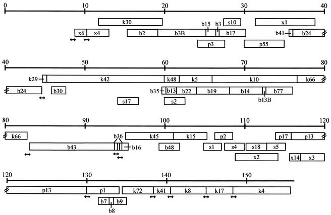
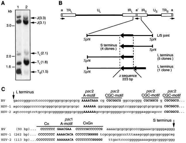
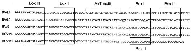
The complete DNA sequence of herpes B virus (Cercopithecine herpesvirus 1) strain E2490, isolated from a rhesus macaque, was determined. The total genome length is 156,789 bp, with 74.5% G+C composition and overall genome organization characteristic of alphaherpesviruses. The first and last residues of the genome were defined by sequencing the cloned genomic termini. There were six origins of DNA replication in the genome due to tandem duplication of both oriL and oriS regions. Seventy-four genes were identified, and sequence homology to proteins known in herpes simplex viruses (HSVs) was observed in all cases but one. The degree of amino acid identity between B virus and HSV proteins ranged from 26.6% (US5) to 87.7% (US15). Unexpectedly, B virus lacked a homolog of the HSV gamma(1)34.5 gene, which encodes a neurovirulence factor. Absence of this gene was verified in two low-passage clinical isolates derived from a rhesus macaque and a zoonotically infected human. This finding suggests that B virus most likely utilizes mechanisms distinct from those of HSV to sustain efficient replication in neuronal cells. Despite the considerable differences in G+C content of the macaque and B virus genes (51% and 74.2%, respectively), codons used by B virus are optimal for the tRNA population of macaque cells. Complete sequence of the B virus genome will certainly facilitate identification of the genetic basis and possible molecular mechanisms of enhanced B virus neurovirulence in humans, which results in an 80% mortality rate following zoonotic infection.
Figures
Similar articles
-
Sequence and genetic arrangement of the U(S) region of the monkey B virus (cercopithecine herpesvirus 1) genome and comparison with the U(S) regions of other primate herpesviruses.J Virol. 2002 Feb;76(3):1516-20. doi: 10.1128/jvi.76.3.1516-1520.2002. J Virol. 2002. PMID: 11773425 Free PMC article.
-
Sequence and genetic arrangement of the UL region of the monkey B virus (Cercopithecine herpesvirus 1) genome and comparison with the UL region of other primate herpesviruses.Arch Virol. 2003 May;148(5):989-97. doi: 10.1007/s00705-003-0011-2. Arch Virol. 2003. PMID: 12721804
-
DNA polymerase gene locus of Cercopithecine herpesvirus 1 is a suitable target for specific and rapid identification of viral infection by PCR technology.Virus Genes. 2005 May;30(3):307-22. doi: 10.1007/s11262-004-6773-0. Virus Genes. 2005. PMID: 15830148
-
The DNA sequence of human herpesvirus-6: structure, coding content, and genome evolution.Virology. 1995 May 10;209(1):29-51. doi: 10.1006/viro.1995.1228. Virology. 1995. PMID: 7747482 Review.
-
Monkey B virus (Cercopithecine herpesvirus 1).Comp Med. 2008 Feb;58(1):11-21. Comp Med. 2008. PMID: 19793452 Free PMC article. Review.
Cited by
-
Specific pathogen free macaque colonies: a review of principles and recent advances for viral testing and colony management.J Med Primatol. 2016 Apr;45(2):55-78. doi: 10.1111/jmp.12209. Epub 2016 Mar 1. J Med Primatol. 2016. PMID: 26932456 Free PMC article. Review.
-
B Virus (Macacine herpesvirus 1) Glycoprotein D Is Functional but Dispensable for Virus Entry into Macaque and Human Skin Cells.J Virol. 2015 May;89(10):5515-24. doi: 10.1128/JVI.03568-14. Epub 2015 Mar 4. J Virol. 2015. PMID: 25740986 Free PMC article.
-
Herpes B virus, macacine herpesvirus 1, breaks simplex virus tradition via major histocompatibility complex class I expression in cells from human and macaque hosts.J Virol. 2012 Dec;86(23):12503-11. doi: 10.1128/JVI.01350-12. Epub 2012 Sep 12. J Virol. 2012. PMID: 22973043 Free PMC article.
-
A chromatin insulator-like element in the herpes simplex virus type 1 latency-associated transcript region binds CCCTC-binding factor and displays enhancer-blocking and silencing activities.J Virol. 2006 Mar;80(5):2358-68. doi: 10.1128/JVI.80.5.2358-2368.2006. J Virol. 2006. PMID: 16474142 Free PMC article.
-
Effectiveness of TRIzol in Inactivating Animal Pathogens.Appl Biosaf. 2023 Dec 1;28(4):230-241. doi: 10.1089/apb.2022.0031. Epub 2023 Dec 5. Appl Biosaf. 2023. PMID: 38090354 Free PMC article. Review.
References
-
- Bennett, A. M., L. Harrington, and D. C. Kelly. 1992. Nucleotide sequence analysis of genes encoding glycoproteins D and J in simian herpes B virus. J. Gen. Virol. 73:2963-2967. - PubMed
-
- Borodovsky, M., and J. McIninch. 1993. Recognition of genes in DNA sequence with ambiguities. Biosystems 30:161-171. - PubMed
-
- Cassady, K. A., M. Gross, G. Y. Gillespie, and B. Roizman. 2002. Second-site mutation outside of the US10-12 domain of Δγ134.5 herpes simplex virus 1 recombinant blocks the shutoff of protein synthesis induced by activated protein kinase R and partially restores neurovirulence. J. Virol. 76:942-949. - PMC - PubMed
Publication types
MeSH terms
Substances
Associated data
- Actions
- Actions
- Actions
Grants and funding
LinkOut - more resources
Full Text Sources
Other Literature Sources
