. 2003 Jun;77(11):6293-304.
doi: 10.1128/jvi.77.11.6293-6304.2003.

Transport of the intracisternal A-type particle Gag polyprotein to the endoplasmic reticulum is mediated by the signal recognition particle

Affiliations

Transport of the intracisternal A-type particle Gag polyprotein to the endoplasmic reticulum is mediated by the signal recognition particle

Frauke Fehrmann et al. J Virol. 2003 Jun.

Abstract

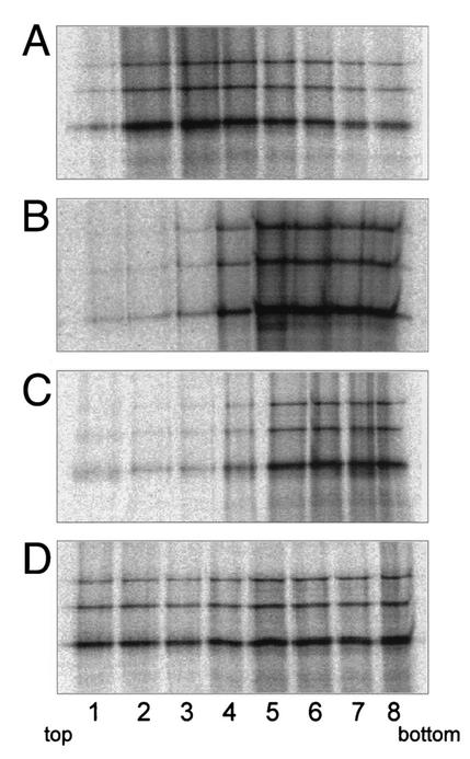
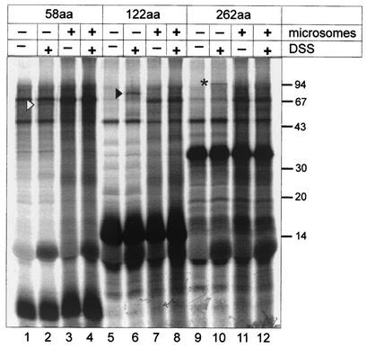
Intracisternal A-type particles (IAP) are defective endogenous retroviruses that accumulate in the endoplasmic reticulum (ER) of rodent cells. The enveloped particles are produced by assembly and budding of IAP Gag polyproteins at the ER membrane. In this study, we analyzed the specific ER transport of the Gag polyprotein of the IAP element MIA14. To this end, we performed in vitro translation of Gag in the presence of microsomal membranes or synthetic proteoliposomes followed by membrane sedimentation or flotation. ER binding of IAP Gag occurred mostly cotranslationally, and Gag polyproteins interacted specifically with proteoliposomes containing only signal recognition particle (SRP) receptor and the Sec61p complex, which form the minimal ER translocation apparatus. The direct participation of SRP in ER targeting of IAP Gag was demonstrated in cross-linking and immunoprecipitation experiments. The IAP polyprotein was not translocated into the ER; it was found to be tightly associated with the cytoplasmic side of the ER membrane but did not behave as an integral membrane protein. Substituting the functional signal peptide of preprolactin for the hydrophobic sequence at the N terminus of IAP Gag also did not result in translocation of the chimeric protein into the ER lumen, and grafting the IAP hydrophobic sequence onto preprolactin failed to yield luminal transport as well. These results suggest that the N-terminal hydrophobic region of the IAP Gag polyprotein functions as a transport signal which mediates SRP-dependent ER targeting, but polyprotein translocation or integration into the membrane is prevented by the signal sequence itself and by additional regions of Gag.


Figures

FIG. 1.
FIG. 1.
(A) Map of the IAP-specific sequence in plasmids pTM1-MIA2, pTM1-MIA4, and pTM1-MIA5. MIA2 corresponds to the wild-type sequence of the IAP MIA14 (38). In MIA4, the first 10 codons of src were substituted for the first 28 codons of the gag gene, and in MIA5 the first 28 codons of gag were deleted. Nucleotide numbering is according to the published sequence (38). Proteins derived from different reading frames are depicted in different lanes. (B) Analysis of in vitro-translated and membrane-bound IAP polyproteins. Coupled transcription-translation reactions were programmed with pTM1-MIA2, pTM1-MIA4, or pTM1-MIA5 in the absence or presence of microsomal membranes as indicated and subsequently centrifuged for 15 min at 12,000 × g. Supernatants (S) and resuspended pellets (P) were applied in equivalent amounts and analyzed by SDS-PAGE. Translation products were labeled with [35S]methionine and detected by autoradiography. The positions of the Gag, Gag-PR, and Gag-PR-Pol polyproteins are indicated on the left. (C) Analysis of membrane-bound IAP polyproteins by sucrose flotation. Coupled transcription-translation reactions were programmed with pTM1-MIA2 or pTM1-MIA5 in the absence or presence of microsomal membranes as indicated. After translation, the samples were adjusted to 85% (wt/vol) sucrose and overlaid with 65 and 10% sucrose. The step gradient was centrifuged at 43,000 rpm in a Beckman Optima TLX ultracentrifuge for 18 h at 4°C. Fractions were collected from top (fraction 1) to bottom (fraction 8), and proteins were resolved by SDS-PAGE.
FIG. 2.
FIG. 2.
Analysis of co- and posttranslational binding of IAP polyproteins. Coupled transcription-translation reactions were programmed with pTM1-MIA2 (A) or pTM1-MIA4 (B) in the absence (lanes 1 and 2) or presence (lanes 3 and 4) of microsomal membranes for 1 h. For analysis of posttranslational transport, reactions without membranes were adjusted to 1.25 mM puromycin and microsomal membranes were added for an additional hour (lanes 5 and 6). Subsequently, translation mixtures were centrifuged for 15 min at 12,000 × g and supernatants (odd lanes), and resuspended pellets (even lanes) were applied in equivalent amounts and analyzed by SDS-PAGE. The relative amounts of soluble and pelleted products were calculated by quantifying radioactively labeled proteins using a Fuji BAS 2000 Bioimager and correcting for the background in each lane. The lower panels depict quantitation of the results from three independent experiments.
FIG. 3.
FIG. 3.
Analysis of IAP polyprotein association with liposomes and reconstituted proteoliposomes. Coupled transcription-translation reactions were programmed with pTM1-MIA2 in the presence of microsomal membranes (A), liposomes prepared from synthetic phospholipids without any protein (B), proteoliposomes reconstituted with purified signal peptidase complex (C), or proteoliposomes reconstituted with SRP receptor and Sec61p complex (D). After translation, all mixtures were subjected to sucrose flotation as described in the legend to Fig. 1C. Fractions were collected from top (fraction 1) to bottom (fraction 8), and proteins were resolved by SDS-PAGE and analyzed by autoradiography.
FIG. 4.
FIG. 4.
Cross-linking of ribosome-nascent-chain complexes to SRP components. Coupled transcription-translation reactions were programmed with pTM1-MIA2 linearized with either StuI (lanes 1 to 4), EcoRI (lanes 5 to 8), or NgoMI (lanes 9 to 12) to produce short mRNAs lacking a stop codon. Reactions were performed for 30 min in the absence or presence of microsomal membranes as indicated. Subsequently, ribosome-nascent-chain complexes were centrifuged at 75,000 rpm in a Beckman Optima TLX ultracentrifuge for 30 min at 2°C. Aliquots from the resuspended pellets were subjected to a cross-linking reaction (2 h, 0°C) with the homobifunctional cross-linking reagent DSS as indicated. Molecular mass markers are indicated on the right. The open arrowhead marks the cross-linked product (58aa × unknown protein) of about 60 kDa in lane 2; a solid arrowhead and an asterisk identify the cross-linked products in lanes 6 and 10, respectively. Note that no specific cross-linking products were detected in lanes 4, 8, and 12, where the corresponding reactions translated in the presence of microsomal membranes were applied.
FIG. 5.
FIG. 5.
Immunoprecipitation of cross-linking products derived from the 122-amino-acid MIA2 polyprotein fragment. Coupled transcription-translation and cross-linking reactions were performed as described in the legend to Fig. 4. Labeled products cross-linked to the 54-kDa subunit of SRP were immunoprecipitated using a specific antiserum directed against SRP54 (lanes 3 and 6). The positions of molecular mass markers are indicated on the right.
FIG. 6.
FIG. 6.
(A) Amino acid sequence at the N termini of ppl, MIA2, and the fusion protein SPpplMIA, in which the signal sequence of MIA2 was replaced by the ppl signal peptide. The amino acid sequences are given in single-letter code, and positively (+) and negatively (−) charged amino acid side chains as well as the cleavage site for signal peptidase (gap) are indicated. The domain representing the ppl signal peptide is shadowed, and the domain representing the IAP signal peptide is boxed. (B) ER transport of ppl, wt IAP Gag and the SPpplMIA fusion protein. Proteins were synthesized by coupled in vitro transcription-translation in the presence or absence of microsomal membranes as indicated. ER import of translation products was analyzed by incubation with proteinase K (50 μg/ml) for 1 h at 0°C in the absence or presence of 0.1% Triton X-100 as indicated. Aliquots of each reaction mixture were analyzed by SDS-PAGE (ppl, lanes 1 to 4; MIA2, lanes 5 to 8, SPpplMIA, lanes 9 to 12). The positions of ppl and the cleaved prolactin (pl) are indicated on the left.
FIG. 7.
FIG. 7.
(A) Amino acid sequence at the N termini of ppl, MIA2, and the fusion proteins plMIA28 and plMIA23, in which the signal sequence of ppl was replaced by the first 28 or 23 amino acids of MIA2, respectively. The amino acid sequences are given in single letter code and positively (+) and negatively (−) charged amino acid side chains as well as the cleavage site for signal peptidase (gap) are indicated. The domain representing the IAP signal peptide is boxed. (B) ER transport of ppl, plMIA28, and plMIA23. Proteins were synthesized and protease treated as described in the legend to Fig. 6 (ppl: lanes 1 to 3; plMIA23:lanes 4 to 6; plMIA28: lane 7 to 9).
FIG. 8.
FIG. 8.
Analysis of IAP Gag polyprotein membrane-association. Coupled in vitro transcription-translation reactions were programmed with pTM-MIA2 in the presence of microsomal membranes, and membrane-bound products were collected by centrifugation. Subsequently, membranes were extracted for 1 h at 25°C with buffer only (lanes 1 and 2); with 1 M NaCl (lanes 3 and 4), 50 mM EDTA (lanes 5 and 6), or 0.1% Triton X-100 (lanes 9 and 10); or with 0.1 M carbonate buffer (pH 11.5) for 30 min at 0°C (lanes 7 and 8). Treated samples were centrifuged for 15 min at 12,000 × g and supernatants (odd lanes) and resuspended pellets (even lanes) were applied in equivalent amounts and analyzed by SDS-PAGE and autoradiography. Relative amounts of soluble and membrane-associated products were calculated as described before, and the lower panel depicts quantitation of the results obtained in three independent experiments.

Similar articles

Cited by

References

    1. Adam, S. A., R. S. Marr, and L. Gerace. 1990. Nuclear protein import in permeabilized mammalian cells requires soluble cytoplasmic factors. J. Cell Biol. 111:807-816. - PMC - PubMed
    1. Anderson, D. J., K. E. Mostov, and G. Blobel. 1983. Mechanisms of integration of de novo-synthesized polypeptides into membranes: signal-recognition particle is required for integration into microsomal membranes of calcium ATPase and of lens MP26 but not of cytochrome b5. Proc. Natl. Acad. Sci. USA 80:7249-7253. - PMC - PubMed
    1. Aota, S., T. Gojobori, K. Shigesada, H. Ozeki, and T. Ikemura. 1987. Nucleotide sequence and molecular evolution of mouse retrovirus-like IAP elements. Gene 56:1-12. - PubMed
    1. Blobel, G., and B. Dobberstein. 1975. Transfer to proteins across membranes. II. Reconstitution of functional rough microsomes from heterologous components. J. Cell Biol. 67:852-862. - PMC - PubMed
    1. Bruss, V., and D. Ganem. 1991. The role of envelope proteins in hepatitis B virus assembly. Proc. Natl. Acad. Sci. USA 88:1059-1063. - PMC - PubMed

Publication types

MeSH terms