. 2003 Jun;77(11):6332-40.
doi: 10.1128/jvi.77.11.6332-6340.2003.

Structure of intracellular mature vaccinia virus visualized by in situ atomic force microscopy

Affiliations

Structure of intracellular mature vaccinia virus visualized by in situ atomic force microscopy

A J Malkin et al. J Virol. 2003 Jun.

Abstract

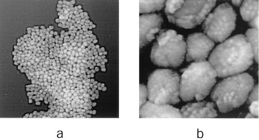
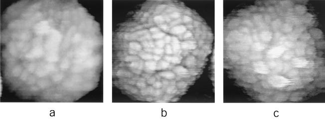
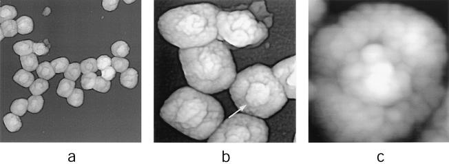
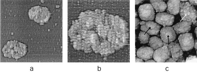
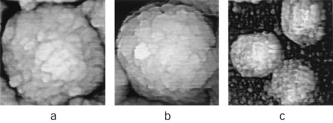
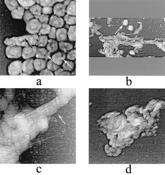
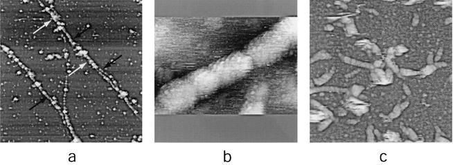
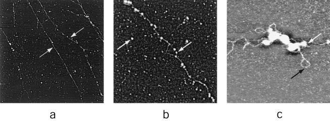
Vaccinia virus, the basis of the smallpox vaccine, is one of the largest viruses to replicate in humans. We have used in situ atomic force microscopy (AFM) to directly visualize fully hydrated, intact intracellular mature vaccinia virus (IMV) virions and chemical and enzymatic treatment products thereof. The latter included virion cores, core-enveloping coats, and core substructures. The isolated coats appeared to be composed of a highly cross-linked protein array. AFM imaging of core substructures indicated association of the linear viral DNA genome with a segmented protein sheath forming an extended approximately 16-nm-diameter filament with helical surface topography; enclosure of this filament within a 30- to 40-nm-diameter tubule which also shows helical topography; and enclosure of the folded, condensed 30- to 40-nm-diameter tubule within the core by a wall covered with peg-like projections. Proteins observed attached to the 30- to 40-nm-diameter tubules may mediate folding and/or compaction of the tubules and/or represent vestiges of the core wall and/or pegs. An accessory "satellite domain" was observed protruding from the intact core. This corresponded in size to isolated 70- to 100-nm-diameter particles that were imaged independently and might represent detached accessory domains. AFM imaging of intact virions indicated that IMV underwent a reversible shrinkage upon dehydration (as much as 2.2- to 2.5-fold in the height dimension), accompanied by topological and topographical changes, including protrusion of the satellite domain. As shown here, the chemical and enzymatic dissection of large, asymmetrical virus particles in combination with in situ AFM provides an informative complement to other structure determination techniques.


Figures

FIG. 1.
FIG. 1.
IMV virions adsorbed to mica. IMV was imaged in situ (in 50 mM Tris-HCl [pH 7.5]). Although isolated virions were observed in all experiments, the vast majority were found, irrespective of the concentration of virions in suspension, to be aggregated into two-dimensional arrays. AFM images are 10 by 10 μm (a) and 1.2 by 1.2 μm (b).
FIG. 2.
FIG. 2.
High-resolution, in situ AFM images of IMV virions (in 50 mM Tris-HCl [pH 7.5]). AFM images are 385 by 385 nm (a), 425 by 425 nm (b), and 365 by 365 nm (c).
FIG. 3.
FIG. 3.
Air-dried IMV virions adsorbed to mica. In panel b the central raised area on the upper surface of one of the virions is indicated with an arrow. AFM images are 2.9 by 2.9 μm (a), 1.0 by 1.0 μm (b), and 350 by 350 nm (c).
FIG. 4.
FIG. 4.
Dissection of IMV virions with nonionic detergent in combination with a reducing agent (1% Igepal and 2% 2-mercaptoethanol in 50 mM Tris-HCl [pH 7.5]; treated for ∼30 to 45 min at 37°C). (a and b) Intact coats. (c) IMV virion cores. The satellite domain structures associated with intact and partially unfolded cores are indicated with black and white arrows, respectively. AFM images are 2.5 by 2.5 μm (a), 1 by 1 μm (b), and 1.35 by 1.35 μm (c).
FIG. 5.
FIG. 5.
High-resolution AFM images of cores after dissection of IMV virions with nonionic detergent in combination with reducing agent (1% Igepal and 2% 2-mercaptoethanol in 50 mM Tris-HCl [pH 7.5]; treated for 30 to 45 min at 37°C). (a and b) Core surface. (c) Seventy- to 100-nm-diameter particles. AFM images are 380 by 380 nm (a), 365 by 365 nm (b), and 400 by 400 nm (c).
FIG. 6.
FIG. 6.
Unfolding of IMV virions. (a to c) Extended treatment with nonionic detergent plus disulfide reducing agent (1% Igepal and 2% 2-mercaptoethanol in 50 mM Tris-HCl [pH 7.5]; treated for 120 to 180 min at 37°C). (a) Several IMV virions (white arrows) have become partially unfolded, resulting in the appearance of 30- to 40-nm-diameter tubules. Other virions (black arrow) have unfolded completely. (b and c) Complete unfolding of IMV results in the appearance of tangled structures formed from 30- to 40-nm-diameter tubules. In panel b intact IMV virions (arrow) are also seen. (d) Thirty- to 40-nm-diameter tubular networks are seen upon treatment of IMV with the reducing agent DTT. AFM images are 1.6 by 1.6 μm (a), 6 by 3 μm (b), 570 by 570 nm (c), and 1.35 by 1.35 μm (d).
FIG. 7.
FIG. 7.
Dissection of IMV virions with proteinase K in combination with ionic detergent. (0.2 to 1 mg of proteinase K per ml in 0.1% sodium dodecyl sulfate-2 mM CaCl2-50 mM HEPES-NaOH [pH 7.5]; treated for ∼45 to 60 min at 37°C). (a) Partially digested portions of 30- to 40-nm-diameter tubules (white arrows) reveal 16-nm-diameter filaments (black arrows). (b and c) Sixteen-nanometer-diameter filaments. AFM images are 6 by 6 μm (a), 240 by 120 nm (b), and 1.5 by 1.5 μm (c).
FIG. 8.
FIG. 8.
DNA strands formed upon extended treatment (90 to 180 min at 37°C) of IMV virions with proteinase K, as in Fig. 7. In panels b and c isolated 16-nm-diameter residual tubular segments are seen dispersed on the mica along with 16-nm-diameter tubular segments associated with DNA strands. Both are indicated with white arrows. In panel c a naked DNA strand is indicated with black arrow. AFM images are 15 by 15 μm (a), 1.5 by 1.5 μm (b), and 580 by 580 nm (c).

References

    1. Baker, T. S., N. H. Olson, and S. D. Fuller. 1999. Adding the third dimension to virus life cycles: three-dimensional reconstruction of icosahedral viruses from cryoelectron micrographs. Microbol. Mol. Biol. Rev. 63:862-922. - PMC - PubMed
    1. Collier, L. 1955. The development of a stable smallpox vaccine. J. Hyg. 53:76-101. - PMC - PubMed
    1. Cudmore, S., R. Blasco, R. Vincentelli, M. Esteban, B. Sodeik, G. Griffiths, and J. K. Locker. 1996. A vaccinia virus core protein, p39, is membrane associated. J. Virol. 70:6909-6921. - PMC - PubMed
    1. Dales, S. 1963. The uptake and development of vaccinia virus in L strain cells followed with labeled viral deoxynucleic acid. J. Cell Biol. 18:51-72. - PMC - PubMed
    1. Dales, S., and B. G. T. Pogo. 1981. Biology of poxviruses. Virology monographs. Springer-Verlag, Vienna, Austria. - PubMed

Publication types