. 2003 Jun;77(11):6567-73.
doi: 10.1128/jvi.77.11.6567-6573.2003.

The herpes simplex virus 1 US3 protein kinase blocks caspase-dependent double cleavage and activation of the proapoptotic protein BAD

Affiliations

The herpes simplex virus 1 US3 protein kinase blocks caspase-dependent double cleavage and activation of the proapoptotic protein BAD

Luca Benetti et al. J Virol. 2003 Jun.

Abstract

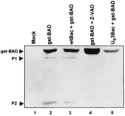
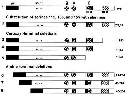
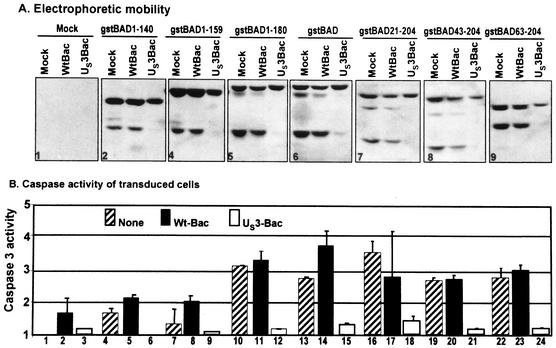
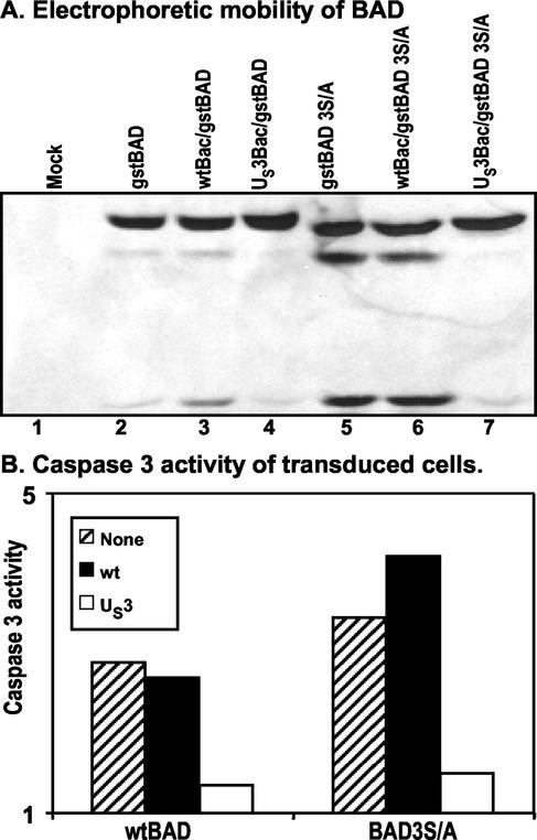
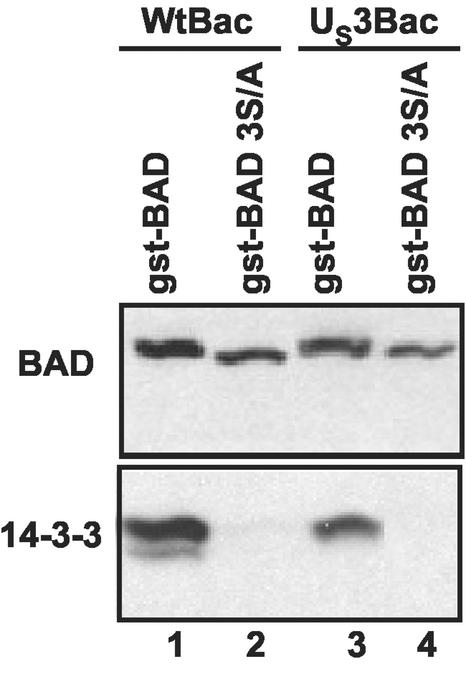
An earlier report showed that the U(S)3 protein kinase blocked the apoptosis induced by the herpes simplex virus 1 (HSV-1) d120 mutant at a premitochondrial stage. Further studies revealed that the kinase also blocks programmed cell death induced by the proapoptotic protein BAD. Here we report the effects of the U(S)3 protein kinase on the function and state of a murine BAD protein. Specifically, (i) in uninfected cells, BAD was processed by at least two proteolytic cleavages that were blocked by a general caspase inhibitor. The untreated transduced cells expressed elevated caspase 3 activity. (ii) In cells cotransduced with the U(S)3 protein kinase, the BAD protein was not cleaved and the caspase 3 activity was not elevated. (iii) Inasmuch as the U(S)3 protein kinase blocked the proapoptotic activity and cleavage of a mutant (BAD3S/A) in which the codons for the regulatory serines at positions 112, 136, and 155 were each replaced with alanine codons, the U(S)3 protein kinase does not act by phosphorylation of these sites nor was the phosphorylation of these sites required for the antiapoptotic function of the U(S)3 protein kinase. (iv) The U(S)3 protein kinase did not enable the binding of the BAD3S/A mutant to the antiapoptotic proteins 14-3-3. Finally, (v) whereas cleavage of BAD at ASP56 and ASP61 has been reported and results in the generation of a more effective proapoptotic protein with an M(r) of 15,000, in this report we also show the existence of a second caspase-dependent cleavage site most likely at the ASP156 that is predicted to inactivate the proapoptotic activity of BAD. We conclude that the primary effect of U(S)3 was to block the caspases that cleave BAD at either residue 56 or 61 predicted to render the protein more proapoptotic or at residue 156, which would inactivate the protein.


Figures

FIG. 1.
FIG. 1.
Photograph of electrophoretically separated cell lysates reacted with a mouse monoclonal antibody to GST. Replicate 25-cm2 flask cultures of rabbit skin cells were either mock infected or infected with 10 PFU of wt-Bac or US3-Bac per cell, incubated for 6 h, and then superinfected with 10 PFU of gstBAD-Bac per cell. One set of culture was exposed to the general caspase inhibitor Z-VAD-fmk, at a concentration of 50 μM, concurrent with gstBAD-Bac infection. Cells were scraped into their medium, pelleted by low-speed centrifugation at 4°C, rinsed twice with phosphate-buffered saline (PBS) A (0.14 mM NaCl, 3 mM KCl, 10 mM Na2HPO4, and 1.5 mM KH2PO4) in the presence of three phosphatase inhibitors (10 mM NaF, 10 mM β-glycerophosphate, 0.1 mM sodium vanadate), and were lysed in radioimmunoprecipitation assay buffer (1% Nonidet P-40, 0.5% sodium deoxycholate, 0.1% sodium dodecyl sulfate [SDS] in PBS A) in the presence of phosphatase inhibitors (10 mM NaF, 10 mM β-glycerophosphate, 0.1 mM sodium vanadate) and protease inhibitors (Complete; Roche) as recommended by the manufacturer. Lysed cells were stored on ice for 10 min before centrifugation at 14,000 rpm for 10 min (in an Eppendorf centrifuge, model 5415 C). The protein concentration of the supernatant fluids was determined with the aid of a Bio-Rad protein assay according to directions provided by the manufacturer. Protein samples denatured in disruption buffer (50 mM Tris [pH 7.0], 2.75% sucrose, 5% 2-mercaptoethanol, 2% SDS) were electrophoretically separated in a 10% denaturing polyacrylamide gel (70 μg of protein per lane), electrically transferred to a nitrocellulose sheet, blocked, and reacted with a mouse monoclonal antibody specific for GST (Santa Cruz). Protein bands were visualized through enhanced chemiluminescence detection according to the instructions of the manufacturer (Amersham Pharmacia). The upper arrowhead points to the intact chimeric protein. Arrowhead P2 points to a faint band consistent with a polypeptide originating from the cleavage of the chimeric protein at amino acid 56 or 61. Arrowhead P1 points to an additional band with an apparent Mr of 42,000. Note that both expression of the US3 protein kinase and treatment with Z-VAD-fmk blocked the appearance of the two rapidly migrating bands. The construction and the transfer plasmid encoding gstBAD were described elsewhere (22). The gstBAD-expressing baculovirus were constructed by cotransfecting the transfer plasmid gstBAD-MTS-1 with Baculogold DNA (Pharmingen) according to the manufacturer's instructions. The baculovirus encoding the US3 protein kinase under the immediate-early cytomegalovirus promoter has been described elsewhere (21).
FIG. 2.
FIG. 2.
Schematic diagram of the structure of the chimeric protein gstBAD (1) and the mutants used in the present study. Regulatory phosphorylation sites at positions 112, 136, and 155 are shown, along with the introduced mutations (S, serine; A, alanine). Stars indicate the two known caspase cleavage sites at residues 56 and 61. gst, GST. Amino-terminal and carboxyl-terminal truncation mutants of BAD were derived from gstBAD-MTS-1 by means of PCR with the primers listed in Table 1. The amplified truncated forms of BAD were inserted into the BamHI/NotI site of gstBAD-MTS-1, in frame with GST. The gstBAD-3S/A-MTS-1 transfer plasmid, in which the codons encoding S112, S136, and S155 were replaced with codons encoding alanines, was constructed as follows. The EcoRI/NotI fragment from gstBAD-MTS-1, encoding the gstBAD fusion protein, was subcloned into the EcoRI/NotI site of pBSK. The serine codons were then replaced with alanine codons with the aid of the QuikChange site-directed mutagenesis kit (Stratagene) according to the manufacturer's instructions. The oligonucleotides used for this purpose were S112A-sense (AGTCGCCACAGTGCGTACCCAGCGGGG), S112A-antisense (CCCCGCTGGGTACGCACTGTGGCGACT), S136A-sense (CGAGGACGCTCGCGTGCGGCTCCCCCC), S136A-antisense (GGGGGGAGCCGCAGCCGAGCGTCCTCG), S155A-sense (CTCCGAAGGATGGCCGATGAGTTTGAGGG), and S155A-antisense (CCCTCAAACTCATCGGCCATCCTTCGGAG). Mutations were confirmed by sequencing, and gstBAD-3S/A was cloned back into the EcoRI/NotI site of MTS-1 The baculoviruses were constructed and propagated as noted in the legend to Fig. 1.
FIG. 3.
FIG. 3.
Effect of the US3 protein kinase expression on cleavage pattern of gstBAD and derivative mutants expressed via recombinant baculovirus (A) and effect of US3 on DEVDase activity induced by gstBAD and derivative mutants (B) in rabbit skin cells. Replicate 25-cm2 flask cultures of rabbit skin cells were either mock infected or infected with 10 PFU of wt-Bac or US3-Bac per cell, incubated for 6 h, and then superinfected with 10 PFU of gstBAD-Bac or each of the truncation mutants per cell. Cells were harvested 18 h after superinfection, solubilized, electrophoretically separated on denaturing gel, electrically transferred to a nitrocellulose sheet, and reacted with mouse monoclonal antibody against gst. Caspase 3 activity in cellular extracts was assayed by using a tetrapeptide conjugated to phenylnitraniline (DEVD-pNA) (BioMol). The cells were scraped, rinsed twice with PBS A, resuspended in 150 μl of lysis solution A (0.1% CHAPS {3-[(3-cholamidopropyl)-dimethylammonio]-1-propanesulfonate}, 50 mM HEPES [pH 7.4], 1 mM dithiothreitol, 0.1 mM EDTA), and incubated on ice for 10 min. Lysates were then centrifuged at 14,000 rpm for 10 min at 4°C. Supernatant fluids were collected, and the protein content was measured with a Bio-Rad protein assay according to directions provided by the manufacturer. Equal amounts of protein were tested for DEVDase activity according to the manufacturer's instructions (BioMol). Chromophore release was quantified by measuring the absorbance at 405 nm with a spectrophotometer after 2 h. The results are expressed as the fold increase in activity compared to mock-infected cells.
FIG. 4.
FIG. 4.
Effect of the US3 protein kinase expression on cleavage pattern of gstBAD and the gstBAD-3S/A mutant expressed via recombinant baculovirus in rabbit skin cells (A) and effect of US3 on DEVDase activity induced by gstBAD and gstBAD-3S/A (B). Replicate 25-cm2 flask cultures of rabbit skin cells were either mock infected or infected with 10 PFU of wt-Bac or US3-Bac per cell, incubated for 6 h, and then superinfected with 10 PFU of gstBAD-Bac or gstBAD-3S/A-Bac per cell. Cells were harvested and electrophoretically separated or assayed for DEVDase activity as described in the legend to Fig. 3.
FIG. 5.
FIG. 5.
Photograph of immunoblot of affinity purified gstBAD or gstBAD-3S/A and coprecipitated 14-3-3 proteins. Replicate 25-cm2 flask cultures of rabbit skin cells were infected with 10 PFU of wt-Bac or US3-Bac per cell, incubated for 6 h, and then superinfected with 10 PFU of either gstBAD-Bac or gstBAD-3S/A-Bac per cell. Cells were harvested 18 h after the superinfection, lysed in 500 μl of lysis buffer B (142.5 mM KCl, 5 mM MgCl2, 10 mM HEPES, 0.25% Nonidet P-40) in the presence of phosphatase and protease inhibitors, and incubated on ice for 10 min. Lysates were pelleted at 14,000 rpm for 10 min at 4°C. The supernatant fluids were collected and reacted overnight with constant agitation at 4°C with 50 μl of a 50% slurry of glutathione beads (Pharmacia Biotech). The beads were pelleted by low-speed centrifugation and rinsed three times with lysis buffer B. The proteins complexed with the pulled-down gstBAD fusion proteins were denatured in disruption buffer (50 mM Tris [pH 7.0], 2.75% sucrose, 5% 2-mercaptoethanol, 2% SDS) and electrophoretically separated in an 11% denaturing polyacrylamide gel. Immunoblotting was done as described in the legend to Fig. 1. The electrophoretically separated proteins were reacted with a rabbit polyclonal antibody specific for BAD (Santa Cruz) or with a rabbit antibody specific for the 14-3-3 proteins (Santa Cruz) and then visualized by enhanced chemiluminescence detection.

References

    1. Adachi, M., and K. Imai. 2002. The proapoptotic BH3-only protein BAD transduces cell death signals independently of its interaction with Bcl-2. Cell Death Differ. 9:1240-1247. - PubMed
    1. Asano, S., T. Honda, F. Goshima, D. Watanabe, Y. Miyake, Y. Sugiura, and Y. Nishiyama. 1999. US3 protein kinase of herpes simplex virus type 2 plays a role in protecting cornel epithelial cells from apoptosis in infected mice. J. Gen. Virol. 80:51-56. - PubMed
    1. Aubert, M., J. O'Toole, and J. A. Blaho. 1999. Induction and prevention of apoptosis in human Hep-2 cells by herpes simplex virus type 1. J. Virol. 73:10359-10370. - PMC - PubMed
    1. Chittenden, T., C. Flemington, A. B. Houghton, R. G. Ebb, G. J. Gallo, B. Elangovan, G. Chinnadurai, and R. J. Lutz. 1995. A conserved domain in Bak, distinct from BH1 and BH2, mediates cell death and protein binding functions. EMBO J. 14:5589-5596. - PMC - PubMed
    1. Condorelli, F., P. Salomoni, S. Cotteret, V. Cesi, S. M. Srinivasula, E. S. Alnemri, and B. Calabretta. 2001. Caspase cleavage enhances the apoptosis-inducing effects of BAD. Mol. Cell. Biol. 21:3025-3036. - PMC - PubMed

Publication types