

Viral internal ribosome entry site structures segregate into two distinct morphologies
- PMID: 12743317
- PMCID: PMC154988
- DOI: 10.1128/jvi.77.11.6574-6579.2003
Viral internal ribosome entry site structures segregate into two distinct morphologies
Abstract
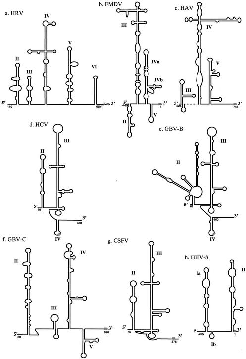
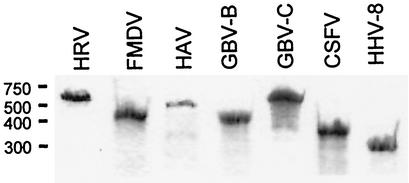
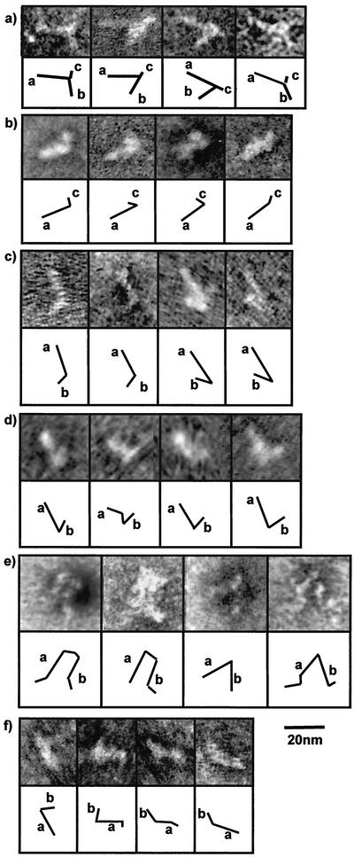
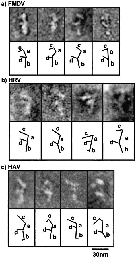
An increasing number of viruses have been shown to initiate protein synthesis by a cap-independent mechanism involving internal ribosome entry sites (IRESs). Predictions of the folding patterns of these RNA motifs have been based primarily on sequence and biochemical analyses. Biophysical confirmation of the models has been achieved only for the IRES of hepatitis C virus (HCV), which adopts an open structure consisting of two major stems. We have conducted an extensive comparison of flavivirus and picornavirus IRES elements by negative stain transmission electron microscopy. All of the flavivirus IRESs we examined (those of GB virus-B, GB virus-C, and classical swine fever virus) fold to give a structure similar to that of the HCV IRES, as does an IRES recently found on mRNA encoded by human herpesvirus 8. The larger picornavirus IRESs (those of foot-and-mouth disease virus, rhinovirus, encephalomyocarditis virus, and hepatitis A virus) are morphologically similar, comprising a backbone with two protruding stems, and distinct from the flavivirus IRESs.
Figures
References
-
- Borman, A., and R. J. Jackson. 1992. Initiation of translation of human rhinovirus RNA: mapping the internal ribosome entry site. Virology 188:685-696. - PubMed
-
- Borman, A. M., and K. M. Kean. 1997. Intact eukaryotic initiation factor 4G is required for hepatitis A virus internal initiation of translation. Virology 237:129-136. - PubMed
Publication types
MeSH terms
Substances
LinkOut - more resources
Full Text Sources
Miscellaneous
