

Review
doi: 10.1136/gut.52.suppl_4.iv12.
Novel methods of enhanced endoscopic imaging
Affiliations
- PMID: 12746263
- PMCID: PMC1867757
- DOI: 10.1136/gut.52.suppl_4.iv12
Item in Clipboard
Review
Novel methods of enhanced endoscopic imaging
Gut.
2003 Jun.
Abstract
Endoscopy has become an essential part of the practice of gastroenterology. Techniques exploiting previously unused properties of light have demonstrated the potential to enhance the ability to make clinical diagnoses without removing tissue as has been standard practice for decades. The term used for many of these techniques is "optical biopsy" and, although not yet widely available, enthusiasm for such techniques has grown as has research in their potential clinical utility.
Figures
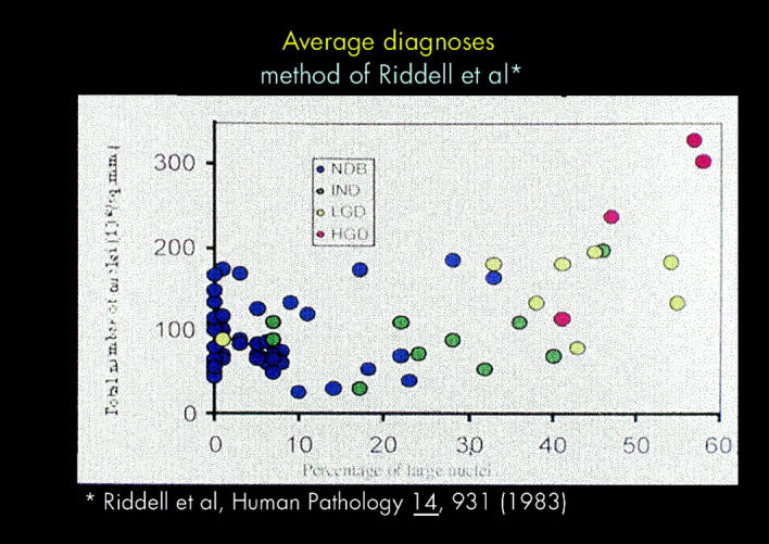
The degree of dysplasia (NDB, non-dysplastic Barrett's; IND, indeterminate for dysplasia; LGD, low grade dysplasia; HGD, high grade dysplasia) in 13 patients with Barrett's oesophagus. Colour diagnosis indicates the average diagnosis of four gastrointestinal pathologists blinded to results obtained by light scattering techniques. Data from reference 12: Gastroenterology 2000;119:677–82.[Medline]
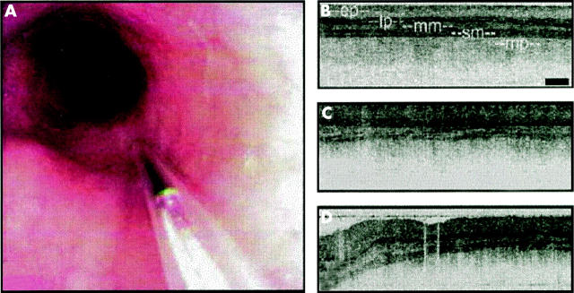
Optical coherence tomography in a patient with a normal oesophagus. (A) endoscopic view of OCT catheter probe in distal oesophagus. (B–D) linear arrangment of mural layers (ep, epitheliaum; lp, lamina propria; mm, muscularis mucosa; sm, submucosa; mp, muscularis propria. From reference 21: Endoscopy 2000;32:921–30.[CrossRef][Medline]
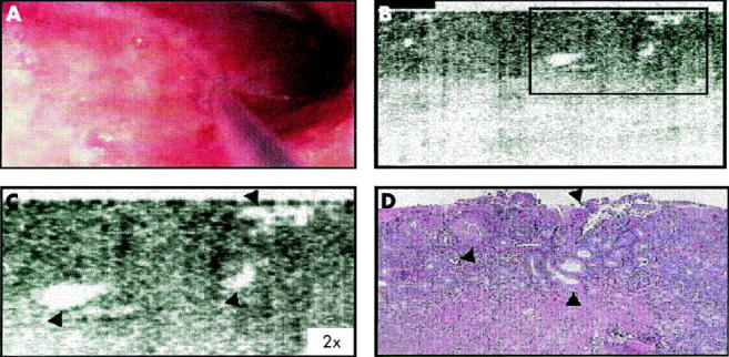
Optical coherence tomography in a patient with Barrett's oesophagus. (A) Endoscopic view of OCT catheter probe in distal oesophagus. (B) OCT image demonstrating loss of linearity and demonstration of submucosal glands. (C) Higher magnification of area in box in B. (D) Histological correlation of OCT image. From reference 21: Endoscopy 2000;32:921–30.[CrossRef][Medline]
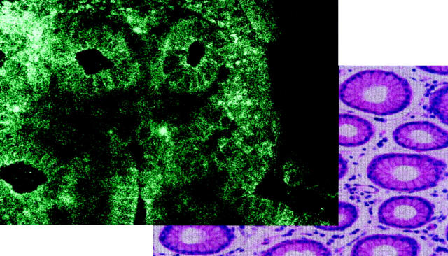
Image of normal gastric mucosal biopsy obtained using confocal microscopy and histological correlate. From reference 28: Endoscopy 2000;32:439–43.[CrossRef][Medline]
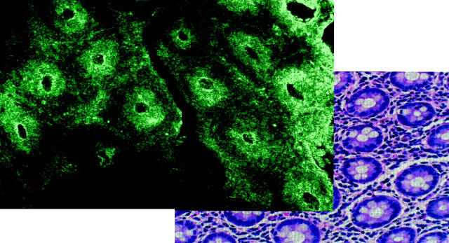
Image of normal colonic mucosal biopsy obtained using confocal microscopy and histological correlate. From reference 28: Endoscopy 2000;32:439–43.[CrossRef][Medline]
References
Publication types
MeSH terms
LinkOut - more resources
Full Text Sources