

Review
doi: 10.1136/gut.52.suppl_4.iv23.
18F-Fluoro-2-deoxyglucose positron emission tomography in the evaluation of gastrointestinal malignancies
Affiliations
- PMID: 12746265
- PMCID: PMC1867761
- DOI: 10.1136/gut.52.suppl_4.iv23
Item in Clipboard
Review
18F-Fluoro-2-deoxyglucose positron emission tomography in the evaluation of gastrointestinal malignancies
Gut.
2003 Jun.
Abstract
Positron emission tomography with (18)F-fluoro-2-deoxyglucose is an imaging technology that is demonstrating increasing utility in the evaluation of gastrointestinal malignancies.
Figures
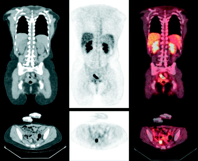
A 45 year old woman with stage III colorectal cancer presented with rising CEA after primary tumour resection. (Top row) Coronal and (bottom row) transaxial tomographic slices. (Left) CT was interpreted as negative for recurrence. (Centre) FDG PET shows intense activity at the anastamosis compatible with tumour recurrence. This was subsequently verified after surgery. (Right) Fusion of PET and CT localises the abnormality.
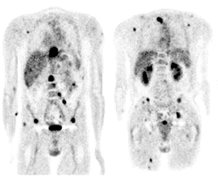
A 57 year old man with oesophageal cancer and clinical stage T3N1 presented for staging evaluation. (Left) Anterior coronal and (right) posterior coronal FDG PET show multiple foci of intense metabolic activity compatible with widespread metastases. CT scan confirmed many of these sites, but was unable to detect spread to lymph nodes and distant soft tissue site.
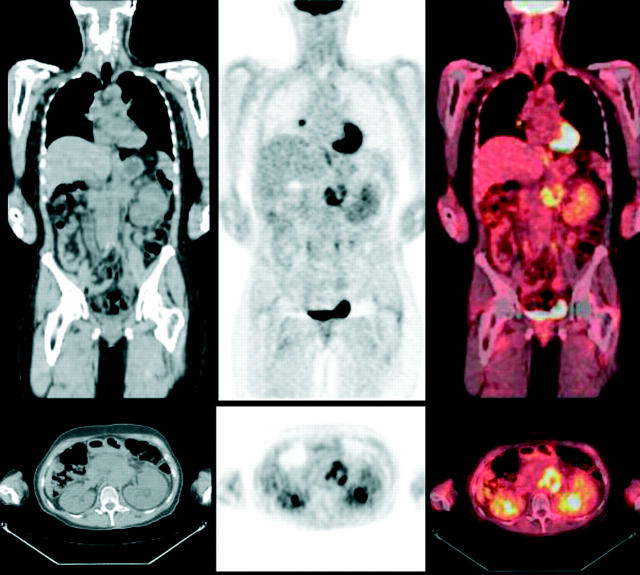
A 43 year old woman with pancreatic cancer presented for evaluation of lung nodules. (Top row) Coronal and (bottom row) transaxial tomographic slices. (Left) CT shows the mass in the abdomen, but is unable to characterise the hilar lymph node. (Centre) FDG PET shows intense activity in the abdominal mass and increased uptake in the hilar lymph node (additional lung abnormalities not shown) compatible with local recurrence and distant metastases. Clinical follow up of lung abnormalities was consistent with metastases. (Right) Fusion of PET and CT localises the abnormalities.
References
Publication types
MeSH terms
Substances
LinkOut - more resources
Full Text Sources