. 2003 Jun 10;100(12):6964-8.
doi: 10.1073/pnas.1030608100. Epub 2003 May 13.

Architecture of initiation-competent 12-subunit RNA polymerase II

Affiliations

Architecture of initiation-competent 12-subunit RNA polymerase II

Karim-Jean Armache et al. Proc Natl Acad Sci U S A. .

Abstract

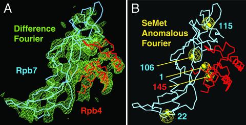
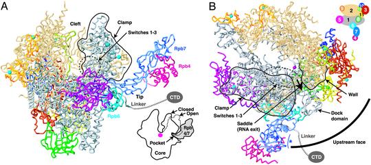
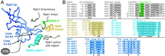
RNA polymerase (Pol) II consists of a 10-polypeptide catalytic core and the two-subunit Rpb4/7 complex that is required for transcription initiation. Previous structures of the Pol II core revealed a "clamp," which binds the DNA template strand via three "switch regions," and a flexible "linker" to the C-terminal repeat domain (CTD). Here we derived a model of the complete Pol II by fitting structures of the core and Rpb4/7 to a 4.2-A crystallographic electron density map. Rpb4/7 protrudes from the polymerase "upstream face," on which initiation factors assemble for promoter DNA loading. Rpb7 forms a wedge between the clamp and the linker, restricting the clamp to a closed position. The wedge allosterically prevents entry of the promoter DNA duplex into the active center cleft and induces in two switch regions a conformation poised for template-strand binding. Interaction of Rpb4/7 with the linker explains Rpb4-mediated recruitment of the CTD phosphatase to the CTD during Pol II recycling. The core-Rpb7 interaction and some functions of Rpb4/7 are apparently conserved in all eukaryotic and archaeal RNA polymerases but not in the bacterial enzyme.


Figures

Fig. 1.
Fig. 1.
Structural analysis of the 12-subunit Pol II. (A) Fit of the Rpb4/7 counterpart structure (15) to the initial 4.2-Å difference Fourier map (green). The map was phased with the Pol II core structure and is contoured at 2σ. Cα backbones for Rpb4 and Rpb7 are shown in pink and blue, respectively. Minor adjustments were made to the structure to account for yeast-specific sequence features. The view corresponds to the “front” view of Pol II (6). (B) Selenomethionine (SeMet) anomalous difference Fourier map (yellow). The Fourier map was calculated with anomalous data from a crystal with selenomethionine-labeled Rpb4/7 (Table 1) and phases from the core model and is contoured at 6σ. Selenomethionine was incorporated as described (52). Five selenium peaks with heights between 20.5σ and 10.6σ coincide with the location of methionine side chains. Indicated as yellow spheres are sulfur atoms in methionine side chains after the corresponding archaeal residues were replaced. No peaks were observed for Rpb4 methionines 1 and 114, which are in flexible regions. The figure was prepared with o (53).
Fig. 2.
Fig. 2.
Architecture of the 12-subunit Pol II, coupling of Rpb4/7 binding and clamp closure, and upstream interaction face. (A) Ribbon model of Pol II. The view is as described for Fig. 1. Cyan spheres and a pink sphere depict eight zinc ions and an active-site magnesium ion, respectively. A black line circles the clamp. The linker to the CTD is indicated as a dashed line. In the lower-right corner, a schematic cut-away view is shown. A dashed line indicates the open clamp position observed in form 2 of the Pol II core structure (7). (B) Pol II upstream interaction face. Shown in a view of the model from the “top” (6). The circle segment is centered at the active site and has a radius that corresponds to the minimal distance between the TATA box and the transcription start site (85 Å, ≈25 bp). The saddle between the wall and the clamp and the assumed direction of RNA exit are indicated. A blue asterisk indicates a potential RNA-binding face of Rpb7 (15, 22). A key to subunit color is shown in the upper right corner. The figure was prepared with RIBBONS (54).
Fig. 3.
Fig. 3.
Pocket–tip interaction. (A) Ribbon model of the Rpb7 tip binding with its two outermost loops (15) to the five protein regions (7) that line the pocket. The view is from the “back,” roughly the reverse of that shown in Figs. 1 and 2A. Colors are as described for Fig. 2 except that switch region 5 is green. (B) Sequence alignments of protein regions are as described for A. Hs, Homo sapiens; Dm, Drosophila melanogaster; Sp, Schizosaccharomyces pombe; Sc, S. cerevisiae; Mj, Methanococcus jannaschii; Ss, Sulfolobus solfataricus. Conserved residues are highlighted. The stars below the alignments indicate invariant residues. The figure was prepared with RIBBONS (54).

Comment in

  • RNA polymerase II at initiation.
    Asturias FJ, Craighead JL. Asturias FJ, et al. Proc Natl Acad Sci U S A. 2003 Jun 10;100(12):6893-5. doi: 10.1073/pnas.1332606100. Epub 2003 Jun 2. Proc Natl Acad Sci U S A. 2003. PMID: 12782794 Free PMC article. No abstract available.

References

    1. Roeder, R. G. (1998) Cold Spring Harbor Symp. Quant. Biol. 63, 201–218. - PubMed
    1. Lee, T. I. & Young, R. A. (2000) Annu. Rev. Genet. 34, 77–137. - PubMed
    1. Lemon, B. & Tjian, R. (2000) Genes Dev. 14, 2551–2569. - PubMed
    1. Ruet, A., Sentenac, A., Fromageot, P., Winsor, B. & Lacroute, F. (1980) J. Biol. Chem. 255, 6450–6455. - PubMed
    1. Edwards, A. M., Kane, C. M., Young, R. A. & Kornberg, R. D. (1991) J. Biol. Chem. 266, 71–75. - PubMed

Publication types

MeSH terms

Associated data