

Apparent diffusion coefficient determination in normal fetal brain: a prenatal MR imaging study
- PMID: 12748074
- PMCID: PMC7975808
Apparent diffusion coefficient determination in normal fetal brain: a prenatal MR imaging study
Abstract
Background and purpose: Diffusion-weighted MR imaging studies of normal brain development have focused on premature babies who were free of focal lesions on conventional MR images. The condition of prematurity, however, is dissimilar to intrauterine life. We sought to establish normal values of fetal brain apparent diffusion coefficient (ADC) to highlight its abnormal changes in pathologic conditions and to obtain information about normal brain development.
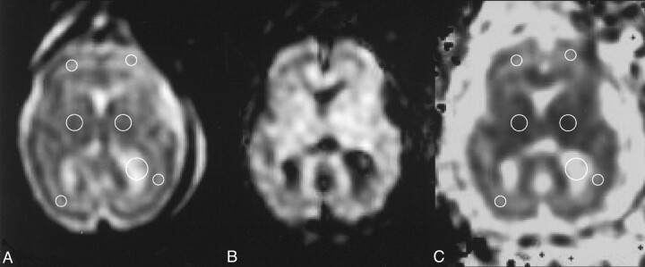
Methods: We measured the ADC, in utero, by using an echo-planar three-axes diffusion-sensitized sequence (b factor, 0 and 600 s/mm(2)), in frontal and occipital white matter and basal ganglia gray matter of 15 fetuses. Their gestational ages ranged from 22 to 35 weeks, and the postnatal MR images or sonograms revealed normal brain.
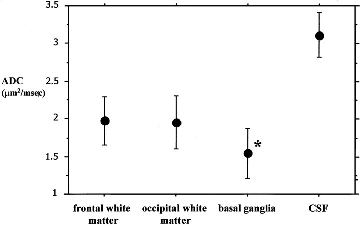
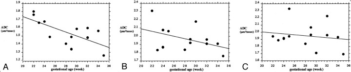
Results: Mean ADC value was 1.96 +/- 0.1 micro m(2)/ms (SD) in frontal white matter, 1.95 +/- 0.1 micro m(2)/ms in occipital white matter, and 1.56 +/- 0.1 micro m(2)/ms in basal ganglia. A significant negative correlation between ADC and gestational age was found for basal ganglia, whereas only a trend was present for frontal white matter.
Conclusion: Although moderately higher, the ADC determinations we obtained are consistent with those reported in the literature in postnatal studies performed in premature babies.
Figures
References
-
- Warach S, Chien D, Li W, et al. Fast magnetic resonance diffusion-weighted imaging of acute human stroke.Neurology 1992;42:1717–1723 - PubMed
-
- Cowan FM, Pennock JM, Hanrahan JD, et al. Early detection of cerebral infarction and hypoxic-ischemic encephalopathy in neonates using diffusion-weighted magnetic resonance imaging.Neuropediatrics 1994;25:172–175 - PubMed
MeSH terms
LinkOut - more resources
Full Text Sources