

The usefulness of MR imaging in the diagnosis of dysembryoplastic neuroepithelial tumor in children: a study of 14 cases
- PMID: 12748079
- PMCID: PMC7975770
The usefulness of MR imaging in the diagnosis of dysembryoplastic neuroepithelial tumor in children: a study of 14 cases
Abstract
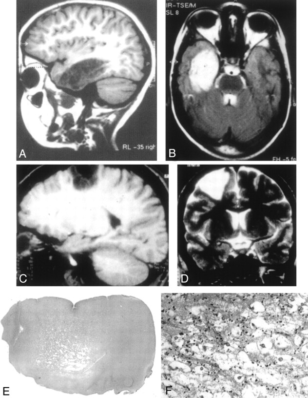
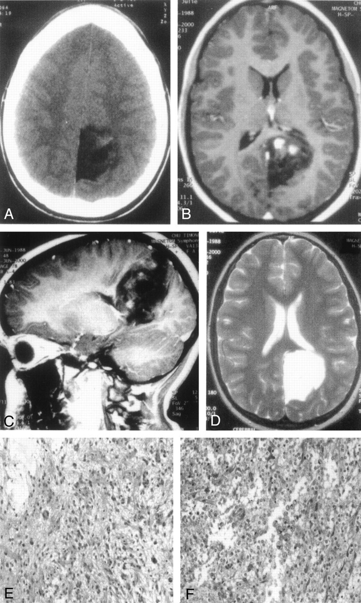
Background and purpose: Dysembryoplastic neuroepithelial tumors (DNTs) are benign lesions affecting children and are associated with epilepsy. The goal of our study was to better characterize the clinical-radiologic-pathologic spectrum of DNTs (complex and simple forms only) in a series of 14 children.
Methods: Clinical, neuroradiologic, and pathologic features of all cases were retrospectively studied.
Results: Eleven cases of complex and three cases of simple DNTs were identified. Mean follow-up was 87 months, and no recurrence was recorded except for one case of simple DNT. We found that some neuroradiologic features may be helpful to support the diagnosis of DNT: presence of "septations," triangular pattern of distribution, and absence of contrast enhancement.
Conclusion: The evidence of the specific glioneuronal element is found by pathologic examination, but the typical neuroradiologic aspect of DNT suggests this diagnosis preoperatively. Radiologic examination may be helpful for the diagnosis of DNT when pathologic findings are inconclusive.
Figures
References
-
- Daumas-Duport C, Scheithauer BW, Chodkiewicz JP, et al. Dysembryoplastic neuroepithelial tumor: a surgically curable tumor of young patients with intractable partial seizures: report of thirty-nine cases. Neurosurgery 1988;23:545–556 - PubMed
-
- Lee DY, Chung CK, Hwang YS, et al. Dysembryoplastic neuroepithelial tumor: radiological findings (including PET, SPECT, and MRS) and surgical strategy. J Neurooncol 1995;47:167–174 - PubMed
-
- Daumas-Duport C, Varlet P, Bacha S, et al. Dysembryoplastic neuroepithelial tumors: nonspecific histological forms: a study of 40 cases. J Neurooncol 1999;41:267–280 - PubMed
-
- Fomekong E, Baylac F, Moret C, et al. Dysembryoplastic neuroepithelial tumors: analysis of 16 cases. Neurochirurgie 1999;45:180–189 - PubMed
-
- Lemesle M, Borsotti JP, Justrabo E, et al. Dysembryoplastic neuroepithelial tumors: a benign tumor cause of partial epilepsy in young adults. Rev Neurol (Paris) 1996;152:451–457 - PubMed
Publication types
MeSH terms
LinkOut - more resources
Full Text Sources
Medical