

Case Reports
Meningocele-induced positional syncope and retinal hemorrhage
Affiliations
- PMID: 12748081
- PMCID: PMC7975804
Item in Clipboard
Case Reports
Meningocele-induced positional syncope and retinal hemorrhage
AJNR Am J Neuroradiol.
2003 May.
Abstract
Meningocele is recognized as a rare, usually asymptomatic condition not associated with acute neurologic symptoms. We herein describe the case of a patient with a longstanding history of a lower back "mass" and recurrent syncope who became acutely unresponsive and developed bilateral retinal hemorrhages when she was placed in the supine position to undergo carotid sonography. MR imaging revealed a large, dorsal lumbar meningocele. The episode likely was caused by acutely increased intracranial pressure caused by displacement of CSF from the meningocele intracranially.
Figures
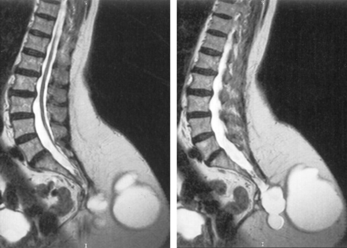
Parasagittal view T2-weighted MR images show the large dorsal meningocele and the tethered cord.
References
-
- Barkovich AJ. Congenital anomalies of the spine. In: Pediatric Neuroimaging. 3rd ed. Philadelphia: Lippincott Williams & Wilkins;2000. :227–271
-
- Naidich TP. The pediatric spine and cord: developmental and congenital abnormalities. In: Categorical Course on Spine and Cord Imaging. Oak Brook: American Society of Neuroradiology;1988. :1–15
-
- Florence G, Seylaz J. Rapid autoregulation of cerebral blood flow: a laser-Doppler flowmetry study. J Cereb Blood Flood Metab 1992;12:674–680 - PubMed
-
- Piper I. Intracranial pressure and elastance. In: Riley P, Bullock R, eds. Head Injury. 1997. :101–120
-
- Brow JK. Mechanisms of production of raised intracranial pressure. In: Minns RA, ed. Problems of Intracranial Pressure in Childhood. London: MacKeith Press;1991. :13–35
Publication types
MeSH terms
LinkOut - more resources
Full Text Sources
Medical