

Focal lesion in the splenium of the corpus callosum on FLAIR MR images: a common finding with aging and after brain radiation therapy
- PMID: 12748085
- PMCID: PMC7975819
Focal lesion in the splenium of the corpus callosum on FLAIR MR images: a common finding with aging and after brain radiation therapy
Abstract
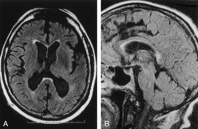
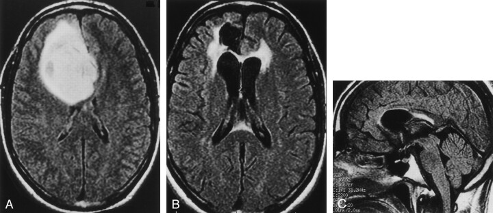
Background and purpose: Focal high signal intensity in the splenium of the corpus callosum on fluid-attenuated inversion-recovery (FLAIR) images is generally considered an abnormal MR finding. We identified high signal intensity in the splenium on FLAIR images in patients of advanced age with otherwise normal images and in patients who had received brain radiation therapy. We undertook an investigation to determine the frequency of this finding in these patient groups.
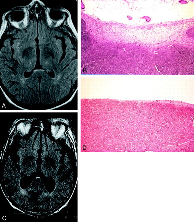
Methods: We reviewed the FLAIR images and medical records of 67 patients (group 1) imaged for suspicion of CNS disease and of 18 consecutive patients (group 2) with history of brain radiation therapy. All FLAIR images were evaluated for focal signal intensity abnormalities in the splenium and for diffuse white matter abnormalities. Also, autopsy specimens from two cases not part of either study group were examined.
Results: Among the initial 67 patients in group 1, focal high signal intensity in the splenium was associated with aging, radiation therapy, and white matter changes. Focal high signal intensity in the splenium was evident on FLAIR images in 16 of the 18 patients in the post-radiation therapy group. Histologic examination of the splenium in one autopsy case with a history of chest and neck radiation therapy demonstrated isomorphic gliosis.
Conclusion: High signal intensity in the splenium of the corpus callosum on FLAIR images is a common finding after brain radiation therapy and can be seen with aging. The radiologist should be aware of this common finding and not mistake it for more commonly recognized causes of splenial lesions.
Figures
Comment in
- AJNR Am J Neuroradiol. 2004 Apr;25(4):664-5
-
Mid-anterior surface of the callosal splenium: subependymal or subpial?AJNR Am J Neuroradiol. 2004 Apr;25(4):664-5. AJNR Am J Neuroradiol. 2004. PMID: 15090365 Free PMC article. No abstract available.
References
-
- Friese SA, Bitzer M, Freudenstein D, et al. Classification of acquired lesion of the corpus callosum with MRI.Neuroradiology 2000;42:795–802 - PubMed
-
- Tsuruda JS, Kortman KE, Bradley WG, et al. Radiation effects on cerebral white matter.AJR Am J Roentgenology 1987;149:165–171 - PubMed
-
- Coffey CE, Wilkinson WE, Parashos IA, et al. Quantitative cerebral anatomy of the aging human brain: a cross-sectional study using magnetic resonance imaging.Neurology 1992;42:527–536 - PubMed
-
- Pantoni L, Garcia JH. Pathogenesis of leukoaraiosis.Stroke 1997;28:652–659 - PubMed
-
- Pantoni L, Garcia JH. The significance of cerebral white matter abnormalities 100 years after Binwanger’s report: a review.Stroke 1995;26:1293–1301 - PubMed
MeSH terms
LinkOut - more resources
Full Text Sources
Medical