

Peritumoral diffusion tensor imaging of high-grade gliomas and metastatic brain tumors
- PMID: 12748097
- PMCID: PMC7975803
Peritumoral diffusion tensor imaging of high-grade gliomas and metastatic brain tumors
Abstract
Background and purpose: Diffusion tensor imaging (DTI) is an advanced MR technique that describes the movement of water molecules by using two metrics, mean diffusivity (MD), and fractional anisotropy (FA), which represent the magnitude and directionality of water diffusion, respectively. We hypothesize that alterations in these values within the tissue surrounding brain tumors reflect combinations of increased water content and tumor infiltration and that these changes can be used to differentiate high-grade gliomas from metastatic lesions.
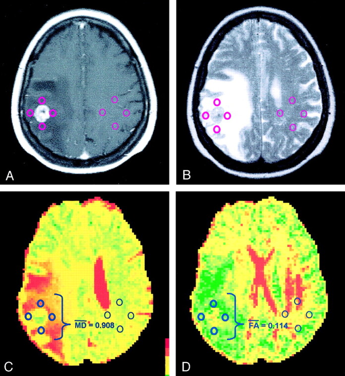
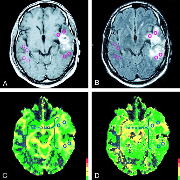
Methods: DTI was performed in 12 patients with high-grade gliomas and in 12 with metastatic lesions. DTI measurements were obtained from regions of interest (ROIs) placed on normal-appearing white matter and on the vasogenic edema, the T2 signal intensity abnormality surrounding each tumor.
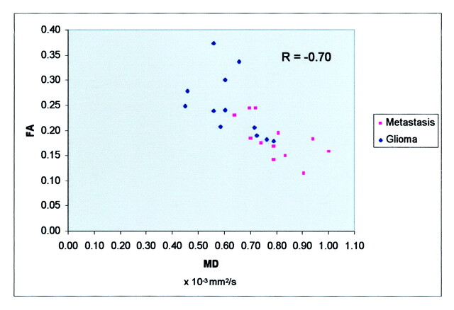
Results: The peritumoral region of both gliomas and metastatic tumors displayed significant increases in MD (P <.005) and significant decreases in FA (P <.005) when compared with those of normal-appearing white matter. Furthermore, the peritumoral MD of metastatic lesions measured significantly greater than that of gliomas (P <.005). Peritumoral FA measurements, on the other hand, showed no such discrepancy.
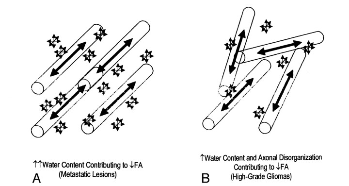
Conclusion: When compared with an internal control, diffusion metrics are clearly altered within the vasogenic edema surrounding both high-grade gliomas and metastatic tumors, reflecting increased extracellular water. Although peritumoral MD can be used to distinguish high-grade gliomas from metastatic tumors, peritumoral FA demonstrated no statistically significant difference. The FA changes surrounding gliomas, therefore, can be attributed not only to increased water content, but also to tumor infiltration.
Figures
References
-
- Alexander AL, Hasan K, Kindlmann G, et al. A geometric analysis of diffusion tensor measurements of the human brain. Magn Res in Med 2000;44:283–291 - PubMed
-
- Bastin ME, Delgado M, Whittle IR, et al. The use of diffusion tensor imaging in quantifying the effect of dexamethasone on brain tumours. NeuroReport 1999;10:1385–1991 - PubMed
-
- Abe O, Aoki S, Hayashi N, et al. Normal aging in the central nervous system: quantitative MR diffusion-tensor analysis. Neurobiol Aging 2002;23:433–441 - PubMed
-
- Krabbe K, Gideon P, Wagn P, et al. MR diffusion imaging of human intracranial tumours. Neurorad 1997;39:483–489 - PubMed
MeSH terms
LinkOut - more resources
Full Text Sources
Medical