

Basilar artery duplication associated with pituitary duplication: a new finding
- PMID: 12748101
- PMCID: PMC7975784
Basilar artery duplication associated with pituitary duplication: a new finding
Abstract
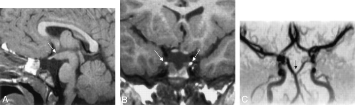
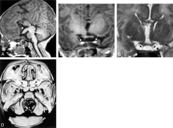
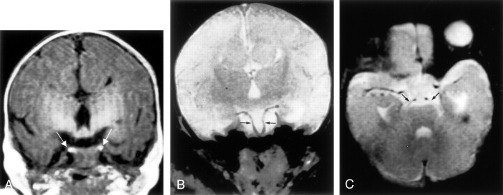
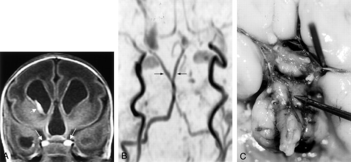
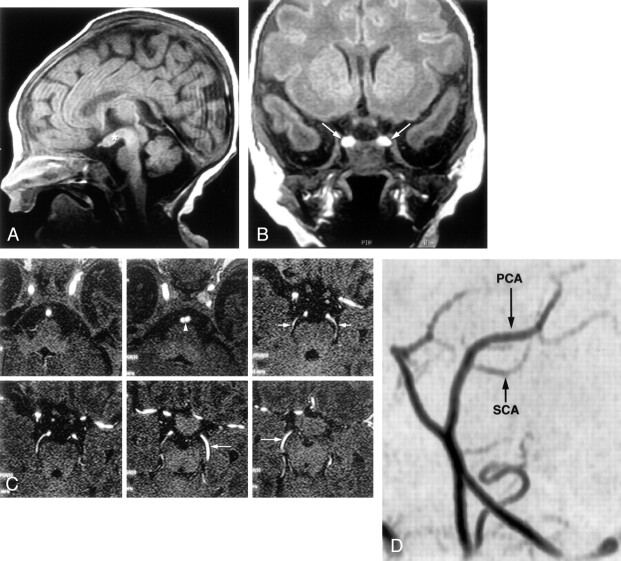
Pituitary duplication is a rare malformation, reported previously in approximately 18 patients. It is usually unsuspected before imaging, although it occurs most commonly in association with complicated midline and skull base anomalies. It is easily shown by MR imaging. Five new cases of pituitary duplication were diagnosed by using MR imaging studies reviewed at the Hospital for Sick Children. Among the many associated midline abnormalities, partial basilar artery duplication is a previously undescribed finding that we observed in all our cases. Cases of basilar artery duplication or fenestration are associated with altered flow dynamics, leading to a higher incidence of aneurysms. Periodic surveillance for this potential complication may be warranted.
Figures
Similar articles
-
Extreme fenestration of the basilar artery associated with cleft palate, nasopharyngeal mature teratoma, and hypophyseal duplication.Eur Radiol. 2002 Aug;12(8):2087-90. doi: 10.1007/s00330-001-1194-0. Epub 2001 Nov 23. Eur Radiol. 2002. PMID: 12136328
-
Duplication of the pituitary gland and basilar artery, with multiple midline fusion defects and craniofacial anomalies.Int J Pediatr Otorhinolaryngol. 2020 Apr;131:109897. doi: 10.1016/j.ijporl.2020.109897. Epub 2020 Jan 21. Int J Pediatr Otorhinolaryngol. 2020. PMID: 31981915
-
Duplication of pituitary gland.J Comput Assist Tomogr. 1997 May-Jun;21(3):459-61. doi: 10.1097/00004728-199705000-00022. J Comput Assist Tomogr. 1997. PMID: 9135658
-
Concurrent basilar artery double fenestration with aneurysm and vertebral artery dissection: case report and literature review of rare cerebrovascular abnormalities.Ann Vasc Surg. 2013 May;27(4):497.e15-21. doi: 10.1016/j.avsg.2012.06.017. Epub 2013 Mar 31. Ann Vasc Surg. 2013. PMID: 23548267 Review.
-
Finding its place on the spectrum of pituitary duplication disorders, duplication of pituitary stalk: A case report with brief review of literature.Neuroradiol J. 2024 Jun;37(3):357-360. doi: 10.1177/19714009231193157. Epub 2023 Jul 28. Neuroradiol J. 2024. PMID: 37507120 Free PMC article. Review.
Cited by
-
Central precocious puberty associated with duplicated pituitary: a case report and literature review.Front Endocrinol (Lausanne). 2025 Feb 11;16:1466411. doi: 10.3389/fendo.2025.1466411. eCollection 2025. Front Endocrinol (Lausanne). 2025. PMID: 40007803 Free PMC article. Review.
-
Endovascular structures of the basilar artery as forms of the basilar nonfusion spectrum.Sci Rep. 2025 Aug 1;15(1):28192. doi: 10.1038/s41598-025-14558-z. Sci Rep. 2025. PMID: 40750985 Free PMC article.
-
Duplication of the pituitary gland - plus syndrome.Indian J Radiol Imaging. 2016 Jan-Mar;26(1):126-30. doi: 10.4103/0971-3026.178361. Indian J Radiol Imaging. 2016. PMID: 27081236 Free PMC article.
-
Hypothalamic hamartoma associated with a craniopharyngeal canal.AJNR Am J Neuroradiol. 2005 Jan;26(1):65-7. AJNR Am J Neuroradiol. 2005. PMID: 15661703 Free PMC article.
-
Pituitary duplication: a rare cause of precocious puberty.Childs Nerv Syst. 2011 Jul;27(7):1157-60. doi: 10.1007/s00381-011-1443-8. Epub 2011 Apr 12. Childs Nerv Syst. 2011. PMID: 21484457 No abstract available.
References
-
- Kollias SS, Ball WS, Prenger EC. Review of the embryologic development of the pituitary gland and report of a case of hypophyseal duplication detected by MRI.Neuroradiology 1995;37:3–12 - PubMed
-
- Shah S, Pereira JK, Becker CJ, Roubal SE. Duplication of pituitary gland.J Comput Assist Tomogr 1997;21:459–561 - PubMed
-
- Hamon-Kerautret M, Ares GS, Demondion X, Rouland V, Francke JP, Pruvo JP. Duplication of the pituitary gland in a newborn with median cleft face syndrome and nasal teratoma.Pediatr Radiol 1998;28:290–292 - PubMed
-
- Lam WW, Metreweli C. MR of double hypophysis.Clin Radiol 1999;54:774–775 - PubMed
-
- Burke M, Zinkovsky S, Abrantes MA, Riley W. Duplication of the hypophysis.Pediatr Neurosurg 2000;33:95–99 - PubMed
Publication types
MeSH terms
LinkOut - more resources
Full Text Sources
Medical