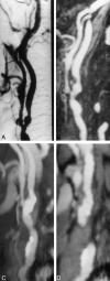
Prospective evaluation of carotid artery stenosis: elliptic centric contrast-enhanced MR angiography and spiral CT angiography compared with digital subtraction angiography
- PMID: 12748115
- PMCID: PMC7975781
Prospective evaluation of carotid artery stenosis: elliptic centric contrast-enhanced MR angiography and spiral CT angiography compared with digital subtraction angiography
Abstract
Background and purpose: Although digital subtraction angiography (DSA) is the reference standard for assessing carotid arteries, it is uncomfortable for patients and has a small risk of disabling stroke and death. These problems have fueled the use of spiral CT angiography and MR angiography. We prospectively compared elliptic centric contrast-enhanced MR angiography and spiral CT angiography with conventional DSA for detecting carotid artery stenosis.
Methods: Eighty carotid arteries (in 40 symptomatic patients) were assessed. Elliptic centric MR and spiral CT angiographic data were reconstructed with maximum intensity projection and multiplanar reconstruction techniques. All patients had been referred for DSA evaluation on the basis of findings at Doppler sonography, which served as a screening method (degree of stenosis > or = 70% or inconclusive results). Degree of carotid stenosis estimated by using the three modalities was compared.
Results: Significant correlation with DSA was found for stenosis degree for both elliptic centric MR and spiral CT angiography; however, the correlation coefficient was higher for MR than for CT angiography (r = 0.98 vs r = 0.86). Underestimation of stenoses of 70-99% occurred in one case with elliptic centric MR angiography (a 70% stenosis was underestimated as 65%) and in nine cases with spiral CT angiography, in comparison to DSA findings. Overestimation occurred in two cases with MR angiography (stenoses of 65-67% were overestimated as 70-75%). With CT, overestimation occurred in seven cases; a stenosis of 60% in one case was overestimated as 70%. Both techniques confirmed the three cases of carotid occlusion. With elliptic centric MR angiography, carotid stenoses of 70% or greater were detected with high sensitivity, 97.1%; specificity, 95.2%; likelihood ratio (LR) for a positive test result, 20.4; and ratio of LR(+) to LR(-), -0.3. With spiral CT angiography, sensitivity, specificity, LR(+), and LR(+):LR(-) were 74.3%, 97.6%, 31.2, and 0.3, respectively.
Conclusion: Elliptic centric contrast-enhanced MR angiography is more accurate than spiral CT angiography to adequately evaluate carotid stenosis. Furthermore, elliptic centric contrast-enhanced MR angiography appears to be adequate to replace conventional DSA in most patients examined.
Figures


Comment in
-
Carotid Artery Stenosis: Competition between CT Angiography and MR Angiography.AJNR Am J Neuroradiol. 2004 Apr;25(4):663-4; author reply 664. AJNR Am J Neuroradiol. 2004. PMID: 15090363 Free PMC article. No abstract available.
References
-
- Seeger MD, Barratt BS, Lawson GA, Klingman N. The relationship between carotid plaque composition, morphology, and neurological symptoms. J Surg Res 1995;58:330–336 - PubMed
-
- North American Symptomatic Carotid Endarterectomy Trial Collaborators. Beneficial effect of carotid endarterectomy in symptomatic patients with high-grade carotid stenosis. N Engl J Med 1991;325:445–453 - PubMed
-
- European Carotid Surgery Trialists’ Collaborative Group. MRC European Carotid Surgery Trial: interim results for symptomatic patients with severe (70–99%) or with mild (0–29%) carotid stenosis.Lancet 1991;337:1235–1243 - PubMed
-
- Barnett HJ, Taylor DW, Eliasziw M, et al. Benefit of carotid endarterectomy in patients with symptomatic moderate or severe stenosis: North American Symptomatic Carotid Endarterectomy Trial Collaborators. N Engl J Med 1998;339:1415–1425 - PubMed
-
- Executive Committee for the Asymptomatic Carotid Atherosclerotic Study. Endarterectomy for asymptomatic carotid artery stenosis. JAMA 1995;273:1421–1428 - PubMed
Publication types
MeSH terms
Substances
LinkOut - more resources
Full Text Sources
