

Coronary calcifications in young patients with first, unheralded myocardial infarction: a risk factor matched analysis by electron beam tomography
- PMID: 12748216
- PMCID: PMC1767690
- DOI: 10.1136/heart.89.6.625
Coronary calcifications in young patients with first, unheralded myocardial infarction: a risk factor matched analysis by electron beam tomography
Abstract
Objective: To compare the presence and extent of coronary calcifications in young patients with first, unheralded acute myocardial infarction with matched controls without a history of coronary artery disease.
Methods: In 102 patients under 60 years of age (19-59 years, mean 41 years; 88% male), electron beam tomography was done 1-14 days after acute myocardial infarction, before any coronary intervention. Coronary calcifications were quantified using the Agatston score. Age related calcium centiles were determined based on the Mayo Clinic "epidemiology of coronary calcification" study, and results were compared with a group of 102 controls without coronary artery disease, matched for sex, age, and risk factors.
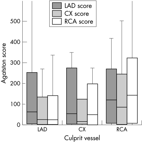
Results: Calcifications were present in 95.1% of patients with acute myocardial infarction and in 59.1% of controls (p = 0.008). The mean (SD) Agatston score was 529 (901) in the infarct patients versus 119 (213) in the controls (p < 0.001). An Agatston score above the 50th centile was present in 87.2% of infarct patients and 47.0% of controls (p = 0.006), and above the 90th centile in 60.7% of infarct patients and only 5.8% of controls (p = 0.001).
Conclusions: In young patients with their first, unheralded acute myocardial infarction, the presence and extent of coronary calcium are significantly greater than in matched controls.
Figures
Similar articles
-
Analysis of coronary calcifications versus Framingham and PROCAM risk assessment in patients with a first myocardial infarction.Int J Cardiol. 2006 Jun 16;110(2):231-6. doi: 10.1016/j.ijcard.2005.09.005. Epub 2005 Nov 28. Int J Cardiol. 2006. PMID: 16310268
-
Predictive value of coronary calcifications for future cardiac events in asymptomatic patients with diabetes mellitus: a prospective study in 716 patients over 8 years.BMC Cardiovasc Disord. 2008 Oct 10;8:27. doi: 10.1186/1471-2261-8-27. BMC Cardiovasc Disord. 2008. PMID: 18847481 Free PMC article.
-
[Coronary heart disease risk in patients without angina pectoris. Coronary calcinosis as a prognostic factor for myocardial infarct?].MMW Fortschr Med. 2001 Apr 19;143(16):27-9. MMW Fortschr Med. 2001. PMID: 11367988 German.
-
[Clinical implication of coronary calcification].Clin Calcium. 2007 Mar;17(3):325-31. Clin Calcium. 2007. PMID: 17339735 Review. Japanese.
-
[Detection and quantification of coronary calcification: an update].Rofo. 2000 May;172(5):407-14. doi: 10.1055/s-2000-666. Rofo. 2000. PMID: 10874967 Review. German.
Cited by
-
Distribution of alkaline phosphatase, osteopontin, RANK ligand and osteoprotegerin in calcified human carotid atheroma.Protein J. 2015 Oct;34(5):315-28. doi: 10.1007/s10930-015-9620-3. Protein J. 2015. PMID: 26307009
-
Noninvasive atherosclerosis imaging for predicting cardiovascular events and assessing therapeutic interventions.Curr Atheroscler Rep. 2004 Jan;6(1):20-6. doi: 10.1007/s11883-004-0112-8. Curr Atheroscler Rep. 2004. PMID: 14662104 Review.
-
Cardiac CT angiography for evaluation of acute chest pain.Int J Cardiovasc Imaging. 2016 Jan;32(1):101-12. doi: 10.1007/s10554-015-0763-2. Epub 2015 Sep 5. Int J Cardiovasc Imaging. 2016. PMID: 26342713 Review.
-
First Report of Massive Myocardial Calcifications in a Vervet Monkey (Chlorocebus pygerythrus).Arch Razi Inst. 2022 Oct 31;77(5):2007-2011. doi: 10.22092/ARI.2022.357062.1963. eCollection 2022 Oct. Arch Razi Inst. 2022. PMID: 37123166 Free PMC article.
-
Visualising noncalcified coronary plaques by CT.Int J Cardiovasc Imaging. 2005 Feb;21(1):55-61. doi: 10.1007/s10554-004-5337-7. Int J Cardiovasc Imaging. 2005. PMID: 15915940 Review.
References
-
- Rumberger JA, Sheedy PF, Breen JF, et al. Electron beam tomography and coronary artery disease: scanning for coronary artery calcification. Mayo Clin Proc 1996;71:369–77. - PubMed
-
- Blankenhorn DH. Coronary artery calcification. Am J Med 1961;31:41–9.
-
- Eggen DA, Strong JP, McGill HC. Coronary calcification: relationship to clinically significant coronary lesions and race, sex and topographic distribution. Circulation 1965;32:948–55. - PubMed
-
- Rumberger JA, Simons DB, Fitzpatrick LA, et al. Coronary artery calcium area by electron beam computed tomography and coronary atherosclerotic plaque area. A histopathologic correlative study. Circulation 1995;92:2157–62. - PubMed
Publication types
MeSH terms
LinkOut - more resources
Full Text Sources
Medical