. 2003 Jun;23(11):3707-20.
doi: 10.1128/MCB.23.11.3707-3720.2003.

Identification of E-box factor TFE3 as a functional partner for the E2F3 transcription factor

Affiliations

Identification of E-box factor TFE3 as a functional partner for the E2F3 transcription factor

Paloma H Giangrande et al. Mol Cell Biol. 2003 Jun.

Abstract

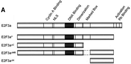
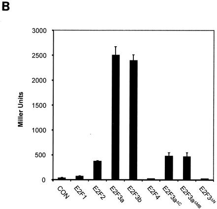
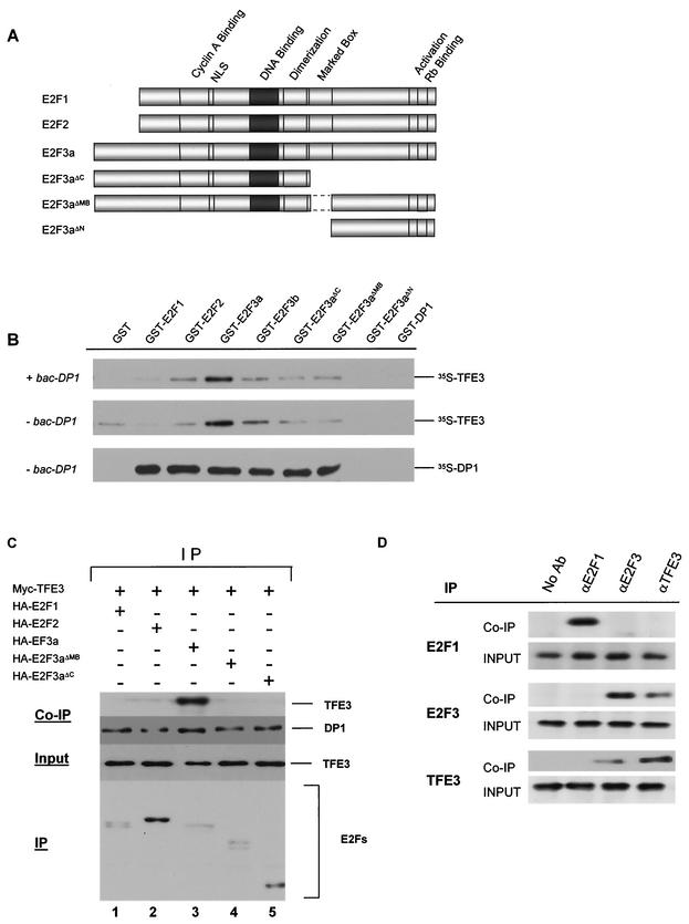
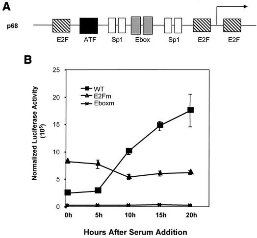
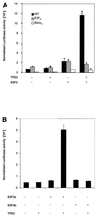
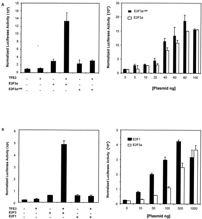
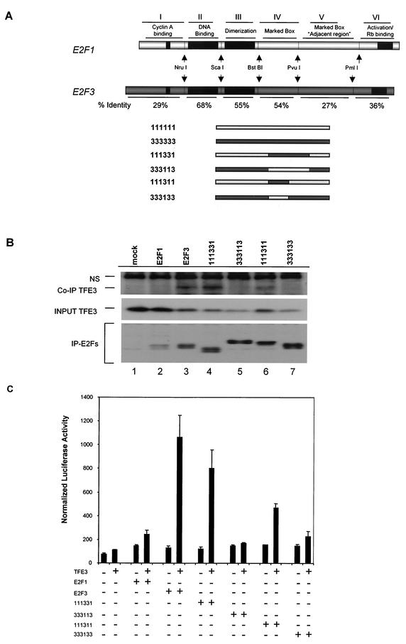
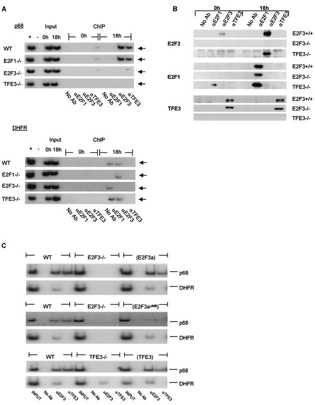
Various studies have demonstrated a role for E2F proteins in the control of transcription of genes involved in DNA replication, cell cycle progression, and cell fate determination. Although it is clear that the functions of the E2F proteins overlap, there is also evidence for specific roles for individual E2F proteins in the control of apoptosis and cell proliferation. Investigating protein interactions that might provide a mechanistic basis for the specificity of E2F function, we identified the E-box binding factor TFE3 as an E2F3-specific partner. We also show that this interaction is dependent on the marked box domain of E2F3. We provide evidence for a role for TFE3 in the synergistic activation of the p68 subunit gene of DNA polymerase alpha together with E2F3, again dependent on the E2F3 marked box domain. Chromatin immunoprecipitation assays showed that TFE3 and E2F3 were bound to the p68 promoter in vivo and that the interaction of either E2F3 or TFE3 with the promoter was facilitated by the presence of both proteins. In contrast, neither E2F1 nor E2F2 interacted with the p68 promoter under these conditions. We propose that the physical interaction of TFE3 and E2F3 facilitates transcriptional activation of the p68 gene and provides strong evidence for the specificity of E2F function.


Figures

FIG. 1.
FIG. 1.
TFE3 is an E2F3-specific binding partner. (A) Schematic of E2F family members and mutants used in the yeast two-hybrid screen. Wild-type E2F1, E2F2, E2F3a, and E2F3b lacking a functional activation domain are designated with a *. E2F3aΔMB has a deletion of the marked box domain (amino acids 295 to 357). In E2F3aΔC the C terminus of E2F3 is deleted (retains amino acids 1 to 295). E2F3aΔN contains amino acids 357 to 398. NLS, nuclear localization signal. (B) A yeast liquid β-galactosidase assay was used to assess the ability of full-length TFE3 to interact with E2F family members E2F1, E2F2, E2F3a, E2F3b, and E2F4 as well as E2F3a deletion mutants. S. cerevisiae was cotransformed with Gal4 AD-TFE3, DP1, and either E2F1, E2F2, EF3a, E2F3b, E2F4, E2F3aΔMB, E2F3aΔN, or E2F3aΔC fused to the Gal4 DBD. Transformants were assayed for β-galactosidase activity, which is represented as Miller units. CON, control (Gal4 DBD).
FIG. 1.
FIG. 1.
TFE3 is an E2F3-specific binding partner. (A) Schematic of E2F family members and mutants used in the yeast two-hybrid screen. Wild-type E2F1, E2F2, E2F3a, and E2F3b lacking a functional activation domain are designated with a *. E2F3aΔMB has a deletion of the marked box domain (amino acids 295 to 357). In E2F3aΔC the C terminus of E2F3 is deleted (retains amino acids 1 to 295). E2F3aΔN contains amino acids 357 to 398. NLS, nuclear localization signal. (B) A yeast liquid β-galactosidase assay was used to assess the ability of full-length TFE3 to interact with E2F family members E2F1, E2F2, E2F3a, E2F3b, and E2F4 as well as E2F3a deletion mutants. S. cerevisiae was cotransformed with Gal4 AD-TFE3, DP1, and either E2F1, E2F2, EF3a, E2F3b, E2F4, E2F3aΔMB, E2F3aΔN, or E2F3aΔC fused to the Gal4 DBD. Transformants were assayed for β-galactosidase activity, which is represented as Miller units. CON, control (Gal4 DBD).
FIG. 2.
FIG. 2.
E2F3 interaction with TFE3. (A) Schematic of E2F family members and mutants used for the in vitro binding assays. Full-length E2F1, E2F2, E2F3a, E2F3b, and E2F3aΔMB have a deletion of the marked box domain (amino acids 295 to 357). In E2F3aΔC the C terminus of E2F3 is deleted (retains amino acids 1 to 295). E2F3aΔN contains amino acids 357 to 398. (B) GST-E2F fusion proteins bound to beads were incubated in the presence of in vitro-translated 35S-labeled Myc-TFE3 or in vitro-translated 35S-labeled DP1 as a control, in either the presence or absence of bacterially expressed DP1. GST alone was used as a negative control in these experiments. (C) NIH 3T3 cells were transfected with Myc-TFE3 and HA-tagged E2F1, E2F2, E2F3a, E2F3aΔMB, or E2F3aΔC (lanes 1 to 5, respectively). Unlike the Gal4 DBD-E2F fusions, the HA-E2F fusions contained a functional activation domain. Transfected cells were lysed in immunoprecipitation lysis buffer at 24 h posttransfection. HA antibody immobilized on Sepharose beads (Covance) was used to immunoprecipitate the HA-tagged E2Fs. Myc-TFE3 and DP1 that coimmunoprecipitated with the E2F proteins were detected by Western blotting with specific antibodies (Co-IP). The level of TFE3 expressed in the transfected cells was measured by Western blotting of aliquots before the immunoprecipitation (input). Finally, the amounts of the E2Fs that were recovered in the immunoprecipitations (IP) was determined by Western blotting with HA antibody. (D) NIH 3T3 cells were lysed in immunoprecipitation lysis buffer, and lysates were incubated with antibodies to E2F1, E2F3, or TFE3 immobilized on protein A- plus protein G-agarose beads (Oncogene). The levels of endogenous E2F1, E2F3, and TFE3 were measured by Western blotting of aliquots before the immunoprecipitation (input). Endogenous TFE3 that coimmunoprecipitated with the E2F proteins was detected by Western blotting with a TFE3-specific antibody. Similarly, endogenous E2F proteins that coimmunoprecipitated with TFE3 were detected by Western blotting with antibodies specific to E2F1 and E2F3. No Ab, no antibody.
FIG. 3.
FIG. 3.
Role of TFE3 and E2F3a in p68 promoter activity. (A) Schematic depiction of the DNA polymerase α p68 subunit promoter. Binding sites for known transcription factors are indicated in relation to the start site of transcription. (B) NIH 3T3 cells were transfected with reporter plasmids and starved for 48 h. After starvation, cells were stimulated to grow by the addition of serum to 20%. Samples were taken at the indicated times, and luciferase activity was measured. Luciferase activity was normalized to β-galactosidase activity (normalized luciferase activity). WT, wild-type promoter; E2Fm, E2F mutant promoter; Eboxm, E-box mutant promoter.
FIG. 4.
FIG. 4.
Synergistic activation of p68 promoter by TFE3 and E2F3a (A) NIH 3T3 cells were transfected with either the wild-type p68 promoter (WT), a promoter containing E2F site mutations (E2Fm), or one with mutations in the E-box elements (Eboxm), together with a plasmid expressing TFE3 alone, E2F3a alone, or TFE3 and E2F3a. Cells were harvested 12 h posttransfection and assayed for luciferase activity and β-galactosidase activity. Luciferase activity was normalized to β-galactosidase activity. (B) NIH 3T3 cells were transfected with the p68 promoter and 10 ng of either empty vector, an E2F3a-expressing plasmid, or an E2F3b-expressing plasmid alone or in combination with 100 ng of the TFE3-expressing plasmid. TFE3 alone was used as a control.
FIG. 5.
FIG. 5.
Synergistic activation of the p68 promoter by E2F and TFE3 is marked box-dependent and specific to E2F3a. (A) Left panel. NIH 3T3 cells were transfected with the p68 promoter and 10 ng of either empty vector, an E2F3a-expressing plasmid, or an E2F3aΔMB-expressing plasmid alone or in combination with 100 ng of the TFE3-expressing plasmid. TFE3 alone was used as a control. Right panel. NIH 3T3 cells were transfected with the p68 promoter and increasing amounts of E2F3a or E2F3aΔMB expression plasmids. Cells were then harvested for determination of luciferase and β-galactosidase activity. (B) Left panel. NIH 3T3 cells were transfected either with 10 ng of plasmids expressing E2F3a or E2F1 or with TFE3 in combination with 10 ng of E2F3a or E2F1. TFE3 alone was used as a control. Cells were harvested as above. Right panel. NIH 3T3 cells were transfected with the wild-type p68 promoter and increasing amounts of expression vector for either E2F1 or E2F3a and harvested for determination of luciferase and β-galactosidase activity as above.
FIG. 6.
FIG. 6.
Marked box domain of E2F3 is both necessary and sufficient for binding to TFE3. (A) Schematic of the chimeric E2Fs described in this study. The nomenclature describes the identity of individual domains in the chimeras: first digit, amino terminus/cyclin A binding; second digit, DNA binding domain; third digit, DP1 dimerization domain; fourth digit, marked box; fifth digit, marked box adjacent region; sixth digit, transactivation/Rb binding domain. The resultant chimeric E2F cDNAs were cloned into a cytomegalovirus-HA expression vector. (B) NIH 3T3 cells were transfected with Myc-TFE3 and HA-tagged E2F1 to E2F3 chimeric proteins. Transfected cells were lysed in immunoprecipitation lysis buffer 24 h posttransfection. HA antibody immobilized on Sepharose beads (Covance) was used to immunoprecipitate the HA-tagged E2Fs. Myc-TFE3 that coimmunoprecipitated with the E2F proteins was detected by Western blotting with specific antibodies (Co-IP TFE3). The level of TFE3 expressed in the transfected cells was measured by Western blotting of aliquots before the immunoprecipitation (input TFE3). Finally, the amounts of the E2Fs that were recovered in the immunoprecipitations (IP-E2Fs) were determined by Western blotting with HA antibody. NS, nonspecific. (C) NIH 3T3 cells were transfected with the p68 promoter and 10 ng of either empty vector, expression plasmids for E2F1 or E2F3, or the E2F1 to E2F3 chimeric proteins alone or in combination with 100 ng of the TFE3 expression plasmid. TFE3 alone was used as a control. Cells were then harvested for determination of Renilla luciferase activity.
FIG. 7.
FIG. 7.
Interaction of E2F3 and TFE3 with the p68 promoter following growth stimulation. (A) Chromatin immunoprecipitation (ChIP) assays were used to examine the interaction of E2F3 and TFE3 with the p68 promoter in intact cells. Mouse embryo fibroblasts (wild type [WT], E2F1−/−, E2F3−/−, and TFE3−/−) were harvested either at quiescence or 18 h following serum stimulation and cross-linked with the addition of formaldehyde as described in the text. Chromatin was immunoprecipitated with antibodies to either E2F1, E2F3, or TFE3. After reversing the cross-link, DNA released from the immunoprecipitates was used for PCR analysis to measure the presence of p68 promoter sequences. The arrows indicate the position of the p68 promoter PCR product. No Ab, no antibody. (B) Aliquots of the immunoprecipitates from panel A were analyzed in an SDS-acrylamide gel and assayed for the presence of E2F1, E2F3, and TFE3 by Western blotting with specific antibodies. (C) Chromatin immunoprecipitation assays were used to examine the interaction of E2F1, E2F3, and TFE3 with the DHFR promoter in intact cells as described above. (D) Chromatin immunoprecipitation assay for interaction of E2F3 and TFE3 with the endogenous p68 and DHFR promoters in wild-type, TFE3−/−, and TFE3−/− MEFs stably expressing TFE3 protein (TFE3).

Similar articles

Cited by

References

    1. Babb, R., C. C. Huang, D. J. Aufiero, and W. Herr. 2001. DNA recognition by the herpes simplex virus transactivator VP16: a novel DNA-binding structure. Mol. Cell. Biol. 21:4700-4712. - PMC - PubMed
    1. Bates, S., A. C. Phillips, P. A. Clark, F. Stott, G. Peters, R. L. Ludwig, and K. H. Vousden. 1998. p14ARF links the tumour suppressors RB and p53. Nature 395:124-125. - PubMed
    1. Brodin, G., A. Hgren, C.-H. Dijke, and R. Heuchel. 2000. Efficient TGF-β induction of the Smad7 gene requires cooperation between AP-1, Sp1, and Smad proteins on the mouse Smad7 promoter. J. Biol. Chem. 275:29023-29030. - PubMed
    1. Cartwright, P., H. Muller, C. Wagener, K. Holm, and K. Helin. 1998. E2F-6: a novel member of the E2F family is an inhibitor of E2F-dependent transcription. Oncogene 17:611-623. - PubMed
    1. Dahl, R., B. Wani, and M. J. Hayman. 1998. The Ski oncoprotein interacts with Skip, the human homolog of Drosophila Bx42. Oncogene 26:1579-1586. - PubMed

Publication types

MeSH terms

LinkOut - more resources