. 2003 Jun;23(11):3951-64.
doi: 10.1128/MCB.23.11.3951-3964.2003.

Activin induces x-zone apoptosis that inhibits luteinizing hormone-dependent adrenocortical tumor formation in inhibin-deficient mice

Affiliations

Activin induces x-zone apoptosis that inhibits luteinizing hormone-dependent adrenocortical tumor formation in inhibin-deficient mice

Felix Beuschlein et al. Mol Cell Biol. 2003 Jun.

Abstract

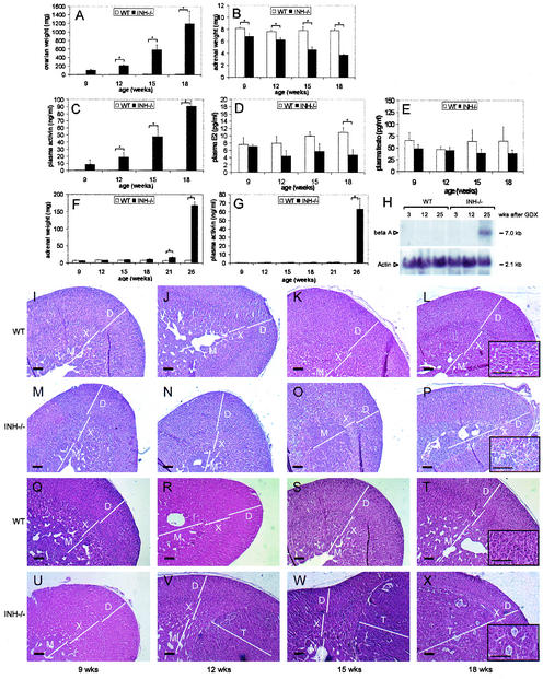
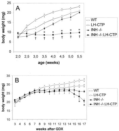
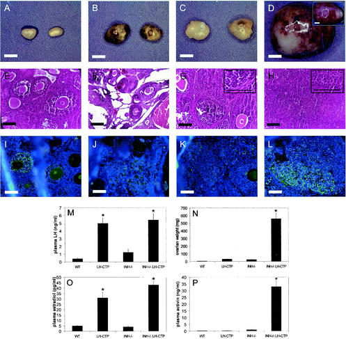
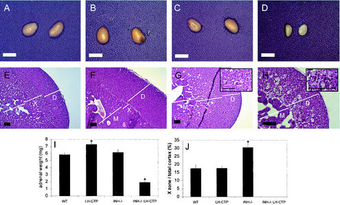
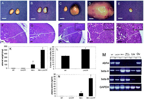
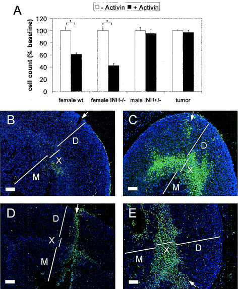
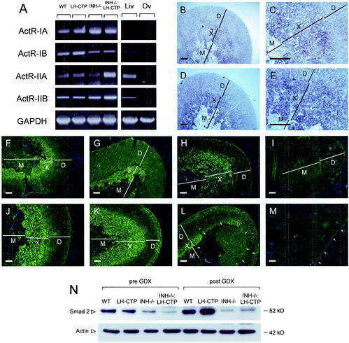
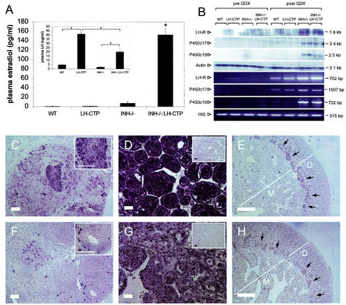
Inhibin and activin are members of the transforming growth factor beta (TGF-beta) family of ligands produced and secreted primarily by the gonads and adrenals. Inhibin-null (INH(-/-)) mice develop gonadal tumors and-when gonadectomized-adrenocortical carcinoma. The mechanisms leading to adrenal tumorigenesis have been proposed to involve the lack of a gonadal factor and/or a compensatory increase in gonadotropins. In order to achieve elevation of gonadotropins without the concomitant loss of a gonadal hormone, we crossed INH(-/-) mice with a transgenic mouse strain that has chronically elevated luteinizing hormone (LH) levels (LH-CTP). Compound INH(-/-)-LH-CTP mice die within 6 weeks of age from severe cancer cachexia induced by large, activin-secreting ovarian tumors. Unexpectedly, INH(-/-)-LH-CTP mice not only fail to develop adrenal tumors but have smaller adrenals, with a regressed x zone, indicating that elevated LH levels are not sufficient to induce adrenal tumor formation. However, following gonadectomy, INH(-/-)-LH-CTP mice develop large, sex steroid-producing adrenal tumors that arise from the x zone, indicating a growth-promoting effect of high levels of LH on the adrenal cortex in the absence of ovarian tumors. In addition, in vivo and in vitro data indicate that activin induces apoptosis specifically in the adrenal x zone. The restricted expression of activin receptor subunits and Smad2 in cells of the adrenal x zone, together with the elevated activin levels in INH(-/-)-LH-CTP mice, supports the conclusion that activin inhibits adrenal tumor growth by inducing x-zone regression.


Figures

FIG. 1.
FIG. 1.
Time course of ovarian and adrenal phenotype in nongonadectomized and gonadectomized wild-type (WT) and INH−/− animals. The expected increase in ovarian tumor weight in INH−/− mice (A) was accompanied by an increase in plasma activin levels (C) and a decrease in adrenal weight (B). Estradiol (D) and testosterone (E) were not elevated in the presence of ovarian tumors. Morphological examination of wild-type (I to L) and INH−/− adrenals (M to P) at 9 (I and M), 12 (J and N), 15 (K and O), and 18 (L and P) weeks of age revealed x-zone regression in INH−/− mice in the presence of ovarian tumors (O and P). When gonadectomized at 6 weeks of age, INH−/− mice developed adrenal tumors with a significant increase in adrenal weight at 21 weeks of age and thereafter (F). Adrenal tumor development was associated with a significant increase in plasma activin levels (G) and expression of activin beta A subunit mRNA in late-stage adrenal tumors (H). Adrenal morphology in gonadectomized wild-type (Q to T) and INH−/− (U to X) mice at 9 (Q and U), 12 (R and V), 15 (S and W), and 18 (T and X) weeks of age revealed a prominent x zone in the 9-week-old INH−/− mice (U) and, consecutively, development of well-circumscribed x-zone tumors (V to X) surrounded by a morphologically intact zona glomerulosa and fasciculata. The inserts in panels L, P, T, and X represent high magnifications of the x-zone area or x-zone tumor, respectively. Bars in panels I to X, including high-magnification inserts, 20 μm. D, definitive zone; X, x zone; M, medulla; T, tumor.
FIG. 2.
FIG. 2.
(A) Body weights of nongonadectomized compound INH−/−-LH-CTP mice were significantly lower than those of wild-type (WT) mice at 3 weeks of age (†) and INH−/− at 3.5 weeks of age (*). All INH−/−-LH-CTP mice died of severe cachexia before or at 6 weeks of age. (B) When gonadectomized at 1.5 weeks of age, compound INH−/−-LH-CTP mice started developing cachexia-like symptoms at 14 weeks after gonadectomy (GDX) and had significantly lower body weights than gonadectomized INH−/− mice at 16 weeks of age and thereafter (*).
FIG. 3.
FIG. 3.
Ovarian phenotypes and hormonal profiles of 5-week-old mice. Gross anatomy (A, B, C, and D), H&E-stained sections (E, F, G, and H), and PCNA immunohistochemistry (I, J, K, and L) of ovaries from wild-type (WT) (A, E, and I), LH-CTP (B, F, and J), INH−/− (C, G, and K), and compound INH−/−-LH-CTP animals (D [contralateral ovary inserted], H, and L). Whereas INH−/− mice developed only small ovarian tumors with retained follicular architecture, INH−/−-LH-CTP mice had massive, undifferentiated ovarian tumors with abundant PCNA expression. As expected, the presence of the LH-CTP transgene led to a significant increase in plasma LH (M) and estradiol (O) levels. The significantly higher tumor weight (N) in INH−/−-LH-CTP mice was accompanied by considerably higher plasma activin levels (P). See the text for a full statistical evaluation. Bars for gross anatomy represent 2 mm; bars for histology, including high-magnification inserts, represent 20 μm.
FIG. 4.
FIG. 4.
Gross appearance and morphological characterization of adrenals from 5-week-old mice. Gross anatomy (A, B, C, and D) and H&E-stained sections (E, F, G, and H) from adrenals of wild-type (WT) (A and E), LH-CTP (B and F), INH−/− (C and G), and compound INH−/−-LH-CTP (D and H) mice. Adrenals from compound INH−/−-LH-CTP mice were significantly smaller than those from mice of the other genotypes (I). In addition, morphological examination revealed an almost complete lack of the x zone. In contrast, the x zone of INH−/− mice was significantly larger than that of wild-type and LH-CTP mice (J), indicating that inhibin deficiency alone is not sufficient to induce x-zone regression. See the text for a full statistical evaluation. Inserts in panels G and H represent high magnifications of the x-zone area. Bars for gross anatomy represent 2 mm; bars for histology, including high-magnification inserts, represent 20 μm. Note the higher magnification in panel H. D, definitive zone; X, x zone; M, medulla.
FIG. 5.
FIG. 5.
Gross appearance and morphological characterization of adrenals from mice 17 weeks after gonadectomy. Gross anatomy (A, B, C, D, and E) and H&E-stained sections (F, G, H, I, and J) from adrenals of wild-type (WT) (A and F), LH-CTP (B and G), INH−/− (C and H), and compound INH−/−-LH-CTP (D, I, E, and J) mice. While inhibin deficiency was necessary for the induction of adrenal tumor growth (C and D), the presence of the LH-CTP transgene resulted in a significantly larger x zone (L) and bigger adrenal tumors, respectively. The higher adrenal weights of INH−/− and INH−/−-LH-CTP mice (K) were paralleled by significantly higher plasma activin levels, as measured by ELISA (N), and adrenal activin beta A and beta B subunit expression, as demonstrated by RT-PCR (M). Notably, the contralateral adrenal from the same INH−/−-LH-CTP animal (E) was small and showed a small x zone consisting of large, multinucleated cells (J). See the text for a full statistical evaluation. The inserts in panels H, I, and J represent high magnifications of the x-zone area or x-zone tumor, respectively. Bars for gross anatomy represent 2 mm; bars for histology, including high-magnification inserts, represent 20 μm. Note the higher magnification in panels I and J. D, definitive zone; X, x zone; T, tumor; M, medulla; Liv, liver; Ov, ovary; GAPDH, glyceraldehyde-3-phosphate dehydrogenase.
FIG. 6.
FIG. 6.
Treatment of primary adrenal cell cultures from female wild-type (wt) and INH−/− mice (including x-zone cells) with recombinant activin A led to a significant reduction in cell number, whereas cells from postpubertal male adrenals (with no x zone) and adrenal tumor cells were unaffected (A). TUNEL staining (green; nuclear counterstain in blue) after injection of medium alone (B and D) and activin (C and E) revealed apoptosis after 3 (B and C) and 6 (D and E) h of incubation specifically in the x zone of activin-treated adrenals. White arrows depict injection channels with some adjacent, mechanically induced apoptotic nuclei. Bars for histology represent 20 μm. D, definitive zone; X, x zone; M, medulla.
FIG. 7.
FIG. 7.
RT-PCR panel demonstrating expression of activin receptor subunits (ActR-IA, ActR-IB, ActR-IIA, and ActR-IIB) in adrenals of wild-type (WT), LH-CTP, INH−/−, and compound INH−/−-LH-CTP mice (A). In situ hybridization for both ActR-IA (B, low magnification; C, high magnification of corticomedullary boundary) and ActR-IIB (D and E) revealed the highest expression of both receptors in the x zone. In addition, immunostaining in adrenals of wild-type (F and J), LH-CTP (G and K), INH−/− (H and L), and compound INH−/−-LH-CTP (I and M) mice before (F to I) and after (J to M) gonadectomy revealed strong x-zone-specific staining of Smad2. In contrast, an adrenal tumor in an INH−/− mouse (L, arrows) showed only weak and partial Smad2 staining whereas no specific staining was detectable in a tumor of an INH−/−-LH-CTP mouse (M). Comparable results demonstrating higher Smad2 expression levels in adrenals with a large x zone and low levels in adrenals with no x zone or with adrenal tumors were obtained by immunoblotting (N). Bars represent 20 μm. Liv, liver; Ov, ovary; D, definitive zone; X, x zone; M, medulla; GAPDH, glyceraldehyde-3-phosphate dehydrogenase.
FIG. 8.
FIG. 8.
Adrenal tumors in inhibin-deficient mice are capable of producing estradiol and express LH receptor, P450c17, and P450arom. Pronounced elevation of estradiol levels in gonadectomized INH−/−-LH-CTP mice (A) was accompanied by partial suppression of elevated LH levels (A, insert). Northern blotting and RT-PCR demonstrated the presence of LH receptor, P450c17, and P450arom transcripts in adrenal tumors of inhibin-deficient mice (B; for details, see text). In situ hybridization for LH receptor (C, D, and E) and P450c17 (F, G, and H) revealed expression of LH receptor and P450c17 in adrenal tumors of gonadectomized INH−/− mice (C and F) and more pronounced expression of LH receptor and P450c17 in compound INH−/−-LH-CTP mice (D and G). Adrenals contralateral to the adrenal tumors in INH−/−-LH-CTP mice (E and H) showed staining for LH receptor and P450c17 transcripts specifically in cells of the remaining x-zone cells (black arrows). Control reactions with a sense probe are shown as inserts in panels D and G. Bars represent 20 μm. Note the higher magnification in panels E and H. WT, wild type; GDX, gonadectomy.

Similar articles

Cited by

References

    1. Beuschlein, F., C. Mutch, D. L. Bavers, Y. M. Ulrich-Lai, W. C. Engeland, C. Keegan, and G. D. Hammer. 2002. Steroidogenic factor-1 is essential for compensatory adrenal growth following unilateral adrenalectomy. Endocrinology 143:3122-3135. - PubMed
    1. Billiar, R. B., M. G. Leavitt, P. Smith, E. D. Albrecht, and G. J. Pepe. 1999. Functional capacity of fetal zone cells of the baboon fetal adrenal gland: a major source of α-inhibin. Biol. Reprod. 61:142-146. - PubMed
    1. Chen, Y. G., H. M. Lui, S. L. Lin, J. M. Lee, and S. Y. Ying. 2002. Regulation of cell proliferation, apoptosis, and carcinogenesis by activin. Exp. Biol. Med. (Maywood) 227:75-87. - PubMed
    1. Cipriano, S. C., L. Chen, K. H. Burns, A. Koff, and M. M. Matzuk. 2001. Inhibin and p27 interact to regulate gonadal tumorigenesis. Mol. Endocrinol. 15:985-996. - PubMed
    1. Coerver, K. A., T. K. Woodruff, M. J. Finegold, J. Mather, A. Bradley, and M. M. Matzuk. 1996. Activin signaling through activin receptor type II causes the cachexia-like symptoms in inhibin-deficient mice. Mol. Endocrinol. 10:534-543. - PubMed

Publication types

MeSH terms

LinkOut - more resources