. 2003 Jun;23(11):4000-12.
doi: 10.1128/MCB.23.11.4000-4012.2003.

In vitro import of a nuclearly encoded tRNA into mitochondria of Solanum tuberosum

Affiliations

In vitro import of a nuclearly encoded tRNA into mitochondria of Solanum tuberosum

Ludovic Delage et al. Mol Cell Biol. 2003 Jun.

Abstract

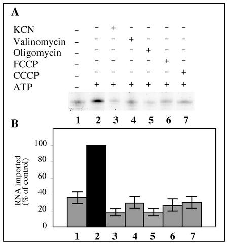
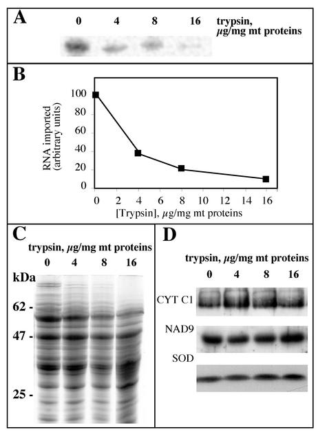
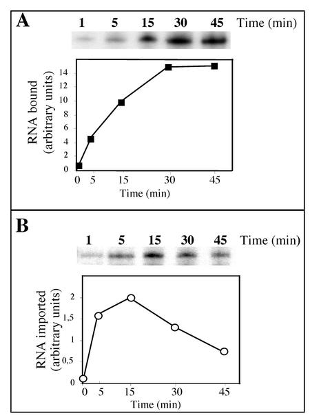
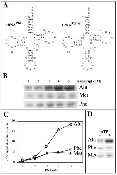
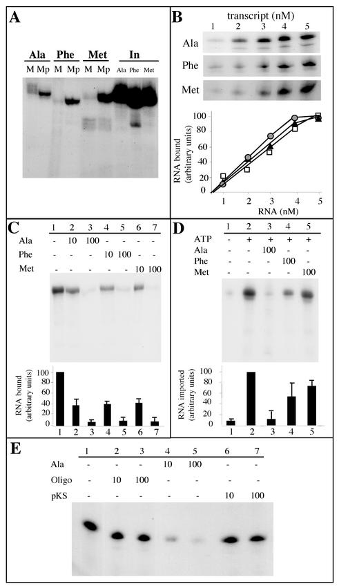
Some of the mitochondrial tRNAs of higher plants are nuclearly encoded and imported into mitochondria. The import of tRNAs encoded in the nucleus has been shown to be essential for proper protein translation within mitochondria of a variety of organisms. Here, we report the development of an in vitro assay for import of nuclearly encoded tRNAs into plant mitochondria. This in vitro system utilizes isolated mitochondria from Solanum tuberosum and synthetic tRNAs transcribed from cloned nuclear tRNA genes. Although incubation of radioactively labeled in vitro-transcribed tRNA(Ala), tRNA(Phe), and tRNA(Met-e) with isolated potato mitochondria resulted in importation, as measured by nuclease protection, the amount of tRNA transcripts protected at saturation was at least five times higher for tRNA(Ala) than for the two other tRNAs. This difference in in vitro saturation levels of import is consistent with the in vivo localization of these tRNAs, since cytosolic tRNA(Ala) is naturally imported into potato mitochondria whereas tRNA(Phe) and tRNA(Met-e) are not. Characterization of in vitro tRNA import requirements indicates that mitochondrial tRNA import proceeds in the absence of any added cytosolic protein fraction, involves at least one protein component on the surface of mitochondria, and requires ATP-dependent step(s) and a membrane potential.


Figures

FIG. 1.
FIG. 1.
The mature tRNAAla is imported in vitro into potato mitochondria. (A) Cloverleaf structure of the mature A. thaliana cytosolic tRNAAla used as a substrate for standard in vitro import assays. (B) Protection of labeled tRNAAla transcripts from RNase degradation. Standard in vitro import assays of 100-μl volumes containing isolated mitochondria corresponding to 400 μg of proteins and 105 cpm of labeled tRNAAla substrate were performed. Reactions were carried out in the presence of 5 mM ATP. After incubation, RNase A and RNase T1 were added to various final concentrations ranging from 0 to 300 μg/ml and from 0 to 1,500 U/ml, respectively. (C) tRNA import is pH dependent. Standard in vitro import assays of 100 μl containing isolated mitochondria corresponding to 400 μg of proteins and 105 cpm of labeled tRNAAla substrate. Reactions were performed in the presence of 5 mM ATP but at different pHs obtained by adjusting the pH of the standard import mixture either with HCl or with NaOH. After import, the pH of all samples was adjusted to 7.2 with HCl or NaOH before addition of RNase A and RNase T1 to final concentrations of 300 μg/ml and 750 U/ml, respectively. Quantification of the amount of protected RNAs was done using a phosphorimager.
FIG. 2.
FIG. 2.
Mitochondrial incorporation of labeled tRNAAla transcripts is ATP dependent and does not require a cytosolic protein fraction. (A) Labeled in vitro-transcribed tRNAAla (105 cpm) was incubated with isolated mitochondria in the presence of increasing concentrations of ATP (0, 2, 5 and 10 mM). In, input RNA (7,500 cpm), no incubation with mitochondria. The import level of the labeled tRNAAla transcripts was quantified using a phosphorimager. (B) Standard in vitro import assays were performed in the presence of 5 mM ATP and in the absence (−) or presence (+) of a bean hypocotyl cytosolic protein extract (PE). (C) Immunodetection of mitochondrial superoxide dismutase (I) and α-tubulin (II) performed with total (T) and mitochondrial (M) extracts from potato tubers.
FIG. 3.
FIG. 3.
The apparent incorporation of tRNAAla in the absence of added ATP is not due to ATP leaking out of mitochondria or to a partial loss of integrity of the mitochondrial membranes but rather to nonspecific protection at the mitochondrial surface. (A) Standard in vitro import assays were performed in the presence of 5 mM ATP (lane 2) and in the presence of apyrase (1 U/ml) (lane 3). After incubation, mild proteinase K digestion and RNase posttreatment were performed as for panel C. (B) Hybridization of a mitochondrial tRNASer-specific oligonucleotide probe to a Northern blot of tRNAs (I) or immunodetection of mitochondrial superoxide dismutase on a Western blot (II). tRNAs and proteins were extracted from the mitochondrial pellet (M) or from the supernatant (S) obtained after centrifugation at 9,000 × g for 10 min of a standard in vitro import assay after 0, 15, and 30 min of incubation. (C) Standard in vitro import assays were performed in the absence (−) or in the presence (+) of 5 mM ATP. After incubation, proteinase K was added to a final concentration of 30 μg/ml, and digestion was carried out for 10 min at room temperature. One hundred microliters of buffer (250 mM mannitol, 10 mM KPO4 [pH 7.5], 0.1% [wt/vol] BSA, 5 mM glycine) containing 2 mM phenylmethylsulfonyl fluoride was added, and the mixture was centrifuged at 15,000 × g for 10 min. RNase posttreatment was then performed as described in Materials and Methods. In, input RNA (1,000 cpm), no incubation with mitochondria.
FIG. 4.
FIG. 4.
Labeled tRNAAla transcript is imported into the matrix of isolated mitochondria. (A) Fractionation and import scheme. (B) Labeled in vitro-transcribed tRNAAla (2 × 10⁵ cpm) was incubated under standard import conditions with intact mitochondria (1); with intact mitochondria, but mitoplasts generated before RNase treatment (2); with mitoplasts (3); with broken mitochondria (4); and without mitochondria (5). (C) The amounts of protected tRNAAla transcript were quantified using a phosphorimager.
FIG. 5.
FIG. 5.
Mitochondrial membrane potential and functional respiration are essential for tRNAAla import. Protonophores (FCCP, lane 6; CCCP, lane 7), an ionophore (valinomycin, lane 4), or specific inhibitors (KCN, lane 3; oligomycin, lane 5) were added to the standard import mixture and incubated for 10 min before adding the labeled tRNAAla transcripts (2 × 10⁵ cpm). As controls, the labeled transcripts were incubated with mitochondria under standard import conditions without (lane 1) or with (lane 2) 5 mM ATP. (B) Histogram showing the import level of the labeled tRNAAla transcript quantified using a phosphorimager. The RNA import level in the presence of ATP and without any inhibitor was taken as 100%.
FIG. 6.
FIG. 6.
In vitro mitochondrial import of tRNAAla transcripts is dependent on surface-accessible proteins. (A) To remove proteins exposed on the outer membrane, isolated mitochondria were treated with increasing concentrations of trypsin (0, 4, 8, and 16 μg of mitochondrial proteins/mg). Labeled in vitro-transcribed tRNAAla (2 × 10⁵ cpm) was then incubated with these different mitochondrial samples under standard import conditions, and protected RNAs were analyzed. (B) The extent of nuclease protection obtained in panel A was quantified using a phosphorimager. (C) Coomassie-blue-stained protein profiles of potato mitochondria treated with increasing concentrations of trypsin as shown in panel A. (D) Corresponding Western blots probed with antibodies directed against CYT C1, NAD9, and SOD.
FIG. 7.
FIG. 7.
Time course of binding or import of tRNAAla transcripts. (A) Kinetics of binding of labeled tRNAAla (105 cpm) onto isolated mitochondria and graphical representation of the results. To test for binding, tRNAAla transcripts were incubated with isolated mitochondria under the same conditions as for tRNA import except that the RNase digestion step was omitted. (B) Kinetics of import of labeled tRNAAla (105 cpm) into isolated mitochondria and graphical representation of the results. Note that between panels A and B, exposure times of the autoradiography were very different (about 10 times longer exposure for import than for binding). For graphical representation of the results, the amounts of bound (squares) or protected (circles) RNAs were quantified using a phosphorimager.
FIG. 8.
FIG. 8.
RNase protection of in vitro-transcribed tRNAAla, tRNAPhe, and tRNAMet incubated with isolated mitochondria. (A) Cloverleaf structures of the mature A. thaliana cytosolic tRNAPhe and tRNAMet-e used as substrates for standard in vitro import assays. (B) Increasing concentrations of labeled tRNAAla, tRNAPhe, and tRNAMet-e were incubated with isolated mitochondria in standard import assays. (C) The amounts of protected RNAs (circles, tRNAAla; squares, tRNAPhe; triangles, tRNAMet-e) were quantified using a phosphorimager, and the graph presented here is the result of three independent experiments. (D) Incubation of 3 nM concentrations of labeled in vitro-transcribed tRNAAla (Ala), tRNAPhe (Phe), and tRNAMet-e (Met) with isolated mitochondria in the absence (−) or in the presence (+) of 5 mM ATP under standard import conditions.
FIG. 9.
FIG. 9.
Analysis of tRNAAla, tRNAPhe, and tRNAMet-e binding to mitochondria or import into mitochondria. (A) Labeled in vitro-transcribed tRNAAla, tRNAPhe, and tRNAMet (2 × 10⁵ cpm each) were incubated under standard import conditions with intact mitochondria (M) or with mitoplasts (Mp). In, input RNAs (5,000 cpm). (B) Bound tRNAAla, tRNAPhe, and tRNAMet-e as a function of tRNAAla, tRNAPhe, and tRNAMet-e concentration, respectively. Increasing concentrations of labeled tRNA transcripts (from 1 to 5 nM) were incubated with isolated mitochondria (corresponding to 100 μg of proteins). Incubation was performed for 10 min under the same conditions as for tRNA import, except that the RNase digestion step was omitted. (C) Competition experiments of tRNAAla binding onto isolated mitochondria using unlabeled tRNAAla, tRNAPhe, or tRNAMet-e as competitors. For these experiments, isolated mitochondria (corresponding to 100 μg of proteins) were preincubated, for 10 min on ice, in the absence of competitor (−) or in the presence of a 10-fold or a 100-fold excess of competitor. Labeled tRNAAla (105 cpm) was then added, and incubation was prolonged for 10 min under the same conditions as for tRNA import, except that the RNase digestion step was omitted. (D) Competition experiments of mitochondrial tRNAAla import into isolated mitochondria using unlabeled tRNAAla, tRNAPhe, or tRNAMet-e as competitor. For these experiments, isolated mitochondria (corresponding to 400 μg of proteins) were preincubated, for 10 min on ice, in the absence of competitor (−) or in the presence of a 100-fold excess of competitor. Then, 2 × 10⁵ cpm of labeled tRNAAla was added, and incubation was prolonged for 15 min under the same conditions as for tRNA import. The experiments were performed in the absence (−) or in the presence (+) of 5 mM ATP. After incubation, a proteinase K treatment was performed as described in Materials and Methods prior to digestion with RNases. (E) Competition experiments of tRNAAla binding to isolated mitochondria using an unlabeled oligodeoxyribonucleotide (Oligo) corresponding to the complete sequence of the cytosol-specific tRNAMet-e (Fig. 8A) or AluI-digested pBluescript vector (pKS) as competitors. The competition experiments were conducted essentially as described for panel C. For graphical representation of the results in panels B, C, and D, the amounts of bound or protected RNAs were quantified using a phosphorimager. Bars represent standard deviations from two and three independent experiments for panels C and D, respectively, and mean values were used to plot the graph.

Similar articles

Cited by

References

    1. Adhya, S., T. Ghosh, A. Das, S. K. Bera, and S. Mahapatra. 1997. Role of an RNA-binding protein in import of tRNA into Leishmania mitochondria. J. Biol. Chem. 272:21396-21402. - PubMed
    1. Akama, K., and M. Kashihara. 1996. Plant nuclear tRNAMet genes are ubiquitously interrupted by introns. Plant Mol. Biol. 32:427-434. - PubMed
    1. Akashi, K., M. Takenaka, S. Yamaoka, Y. Suyama, H. Fukuzawa, and K. Ohyama. 1998. Coexistence of nuclear DNA-encoded tRNAVal (AAC) and mitochondrial DNA-encoded tRNAVal (UAC) in mitochondria of a liverwort Marchantia polymorpha. Nucleic Acids Res. 26:2168-2172. - PMC - PubMed
    1. Aphasizhev, R., U. Karmarkar, and L. Simpson. 1998. Are tRNAs imported into the mitochondria of kinetoplastid protozoa as 5′-extended precursors? Mol. Biochem. Parasitol. 93:73-80. - PubMed
    1. Bauer, M. F., S. Hofmann, W. Neupert, and M. Brunner. 2000. Protein translocation into mitochondria: the role of TIM complexes. Trends Cell Biol. 10:25-31. - PubMed

Publication types

MeSH terms

LinkOut - more resources