

Evolutionary divergence of platelet-derived growth factor alpha receptor signaling mechanisms
- PMID: 12748302
- PMCID: PMC155222
- DOI: 10.1128/MCB.23.11.4013-4025.2003
Evolutionary divergence of platelet-derived growth factor alpha receptor signaling mechanisms
Abstract
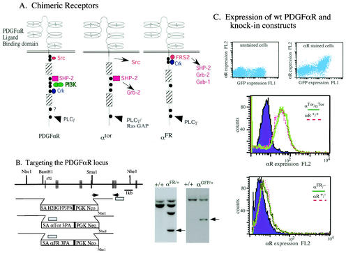
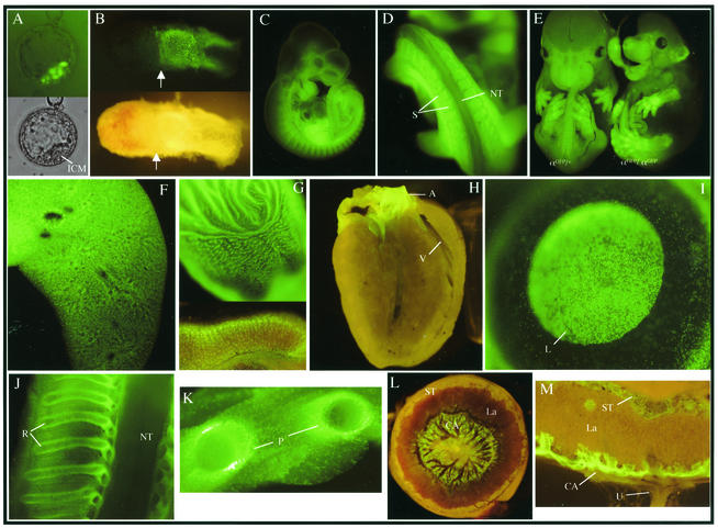
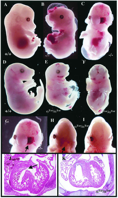
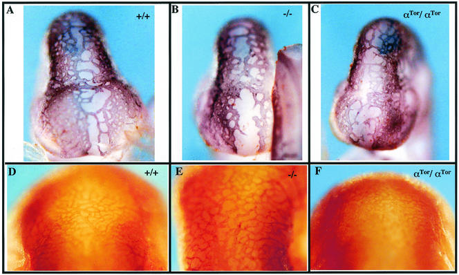
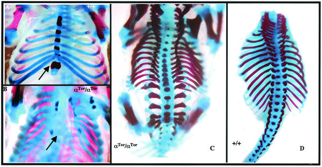
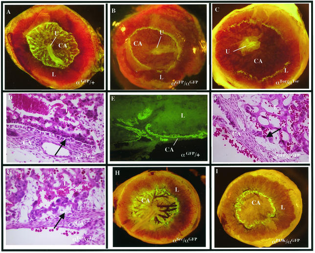
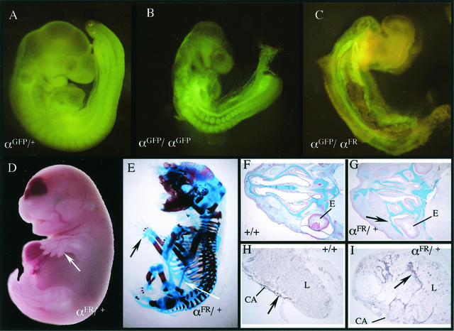
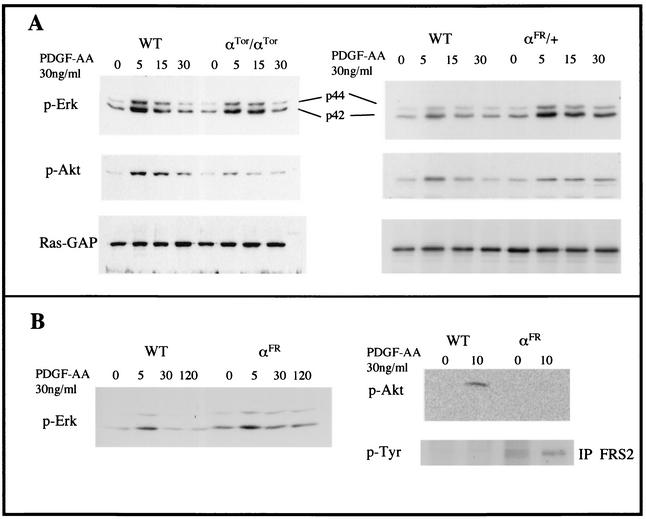
Receptor tyrosine kinases (RTKs) direct diverse cellular and developmental responses by stimulating a relatively small number of overlapping signaling pathways. Specificity may be determined by RTK expression patterns or by differential activation of individual signaling pathways. To address this issue we generated knock-in mice in which the extracellular domain of the mouse platelet-derived growth factor alpha receptor (PDGFalphaR) is fused to the cytosolic domain of Drosophila Torso (alpha(Tor)) or the mouse fibroblast growth factor receptor 1 (alpha(FR)). alpha(Tor) homozygous embryos exhibit significant rescue of neural crest and angiogenesis defects normally found in PDGFalphaR-null embryos yet fail to rescue skeletal or extraembryonic defects. This phenotype was associated with the ability of alpha(Tor) to stimulate the mitogen-activated protein (MAP) kinase pathway to near wild-type levels but failure to completely activate other pathways, such as phosphatidylinositol (PI) 3-kinase. The alpha(FR) chimeric receptor fails to rescue any aspect of the PDGFalphaR-null phenotype. Instead, alpha(FR) expression leads to a gain-of-function phenotype highlighted by ectopic bone development. The alpha(FR) phenotype was associated with a failure to limit MAP kinase signaling and to engage significant PI3-kinase response. These results suggest that precise regulation of divergent downstream signaling pathways is critical for specification of RTK function.
Figures
Similar articles
-
The Shb adaptor protein binds to tyrosine 766 in the FGFR-1 and regulates the Ras/MEK/MAPK pathway via FRS2 phosphorylation in endothelial cells.Mol Biol Cell. 2002 Aug;13(8):2881-93. doi: 10.1091/mbc.e02-02-0103. Mol Biol Cell. 2002. PMID: 12181353 Free PMC article.
-
An allelic series at the PDGFalphaR locus indicates unequal contributions of distinct signaling pathways during development.Dev Cell. 2002 Jan;2(1):103-13. doi: 10.1016/s1534-5807(01)00103-4. Dev Cell. 2002. PMID: 11782318
-
Signaling specificities of fibroblast growth factor receptors in early Xenopus embryo.J Cell Sci. 2000 Aug;113 ( Pt 16):2865-75. doi: 10.1242/jcs.113.16.2865. J Cell Sci. 2000. PMID: 10910771
-
Functions and mechanisms of receptor tyrosine kinase Torso signaling: lessons from Drosophila embryonic terminal development.Dev Dyn. 2005 Mar;232(3):656-72. doi: 10.1002/dvdy.20295. Dev Dyn. 2005. PMID: 15704136 Free PMC article. Review.
-
Receptor Tyrosine Kinases: Translocation Partners in Hematopoietic Disorders.Trends Mol Med. 2017 Jan;23(1):59-79. doi: 10.1016/j.molmed.2016.11.002. Epub 2016 Dec 14. Trends Mol Med. 2017. PMID: 27988109 Review.
Cited by
-
Mapping the molecular and structural specialization of the skin basement membrane for inter-tissue interactions.Nat Commun. 2021 May 10;12(1):2577. doi: 10.1038/s41467-021-22881-y. Nat Commun. 2021. PMID: 33972551 Free PMC article.
-
Dynamic regulation of platelet-derived growth factor receptor α expression in alveolar fibroblasts during realveolarization.Am J Respir Cell Mol Biol. 2012 Oct;47(4):517-27. doi: 10.1165/rcmb.2012-0030OC. Epub 2012 May 31. Am J Respir Cell Mol Biol. 2012. PMID: 22652199 Free PMC article.
-
Hedgehog-responsive mesenchymal clusters direct patterning and emergence of intestinal villi.Proc Natl Acad Sci U S A. 2012 Sep 25;109(39):15817-22. doi: 10.1073/pnas.1205669109. Epub 2012 Sep 10. Proc Natl Acad Sci U S A. 2012. PMID: 23019366 Free PMC article.
-
A novel class of interstitial cells in the mouse and monkey female reproductive tracts.Biol Reprod. 2015 Apr;92(4):102. doi: 10.1095/biolreprod.114.124388. Epub 2015 Mar 18. Biol Reprod. 2015. PMID: 25788664 Free PMC article.
-
Structural and functional properties of platelet-derived growth factor and stem cell factor receptors.Cold Spring Harb Perspect Biol. 2013 Aug 1;5(8):a009100. doi: 10.1101/cshperspect.a009100. Cold Spring Harb Perspect Biol. 2013. PMID: 23906712 Free PMC article. Review.
References
-
- Cleghon, V., P. Feldmann, C. Ghiglione, T. D. Copeland, N. Perrimon, D. A. Hughes, and D. K. Morrison. 1998. Opposing actions of CSW and RasGAP modulate the strength of Torso RTK signaling in the Drosophila terminal pathway. Mol. Cell 2:719-727. - PubMed
-
- Cleghon, V., U. Gayko, T. D. Copeland, L. A. Perkins, N. Perrimon, and D. K. Morrison. 1996. Drosophila terminal structure development is regulated by the compensatory activities of positive and negative phosphotyrosine signaling sites on the Torso RTK. Genes Dev. 10:566-577. - PubMed
-
- Dossenbach, C., S. Rock, and M. Affolter. 2001. Specificity of FGF signaling in cell migration in Drosophila. Development 128:4563-4572. - PubMed
-
- Fambrough, D., K. McClure, A. Kazlauskas, and E. S. Lander. 1999. Diverse signaling pathways activated by growth factor receptors induce broadly overlapping, rather than independent, sets of genes. Cell 97:727-741. - PubMed
Publication types
MeSH terms
Substances
Grants and funding
LinkOut - more resources
Full Text Sources
Other Literature Sources
Molecular Biology Databases
Research Materials
Miscellaneous