

Comment
doi: 10.1172/JCI18611.
A matter of life and death: cardiac myocyte apoptosis and regeneration
Affiliations
- PMID: 12750394
- PMCID: PMC155176
- DOI: 10.1172/JCI18611
Item in Clipboard
Comment
A matter of life and death: cardiac myocyte apoptosis and regeneration
J Clin Invest.
2003 May.
No abstract available
Figures
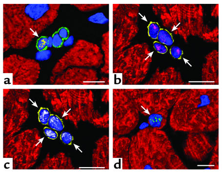
Developing myocytes in a pathologic human heart. (a) Cardiac primitive cells expressing c-kit on the surface membrane (green fluorescence). Nuclei are stained by propidium iodide (blue fluorescence). The nucleus of one of these c-kit–positive cells is labeled by the marker of cell proliferation Ki67 (yellow fluorescence; arrow). (b and c) Four myocyte progenitors illustrated in the same field. They are recognized by the presence of the stem cell surface antigen multi drug resistance protein 1 (MDR-1) (yellow fluorescence; arrows) and by the nuclear localization of the myocyte specific transcription factor myocyte enhancer factor 2 (MEF2) (b, magenta fluorescence). These early committed cells are cycling as documented by the expression of mini chromosome maintenance protein 5 (MCM5) (c, white fluorescence). MCM5 is another marker of the cell cycle. (d) An amplifying myocyte (arrow), which has lost the stem cell surface antigens, is shown. However the nucleus (blue fluorescence) expresses telomerase (green fluorescent dots). A thin layer of myocyte cytoplasm is recognized by the red fluorescence of cardiac myosin heavy chain. Confocal microscopy; scale bars = 10 μm.
Comment on
-
Activation of Mst1 causes dilated cardiomyopathy by stimulating apoptosis without compensatory ventricular myocyte hypertrophy.J Clin Invest. 2003 May;111(10):1463-74. doi: 10.1172/JCI17459. J Clin Invest. 2003. PMID: 12750396 Free PMC article.
-
A mechanistic role for cardiac myocyte apoptosis in heart failure.J Clin Invest. 2003 May;111(10):1497-504. doi: 10.1172/JCI17664. J Clin Invest. 2003. PMID: 12750399 Free PMC article.
References
-
- MacLellan WR, Schneider MD. Genetic dissection of cardiac growth control pathways. Annu. Rev. Physiol. 2000;62:289–319. - PubMed
-
- Kang PM, Izumo S. Apoptosis and heart failure: a critical review of the literature. Circ. Res. 2000;86:1107–1113. - PubMed
-
- Nadal-Ginard B, Kajstura J, Leri A, Anversa P. Myocyte death, growth, and regeneration in cardiac hypertrophy and failure. Circ. Res. 2003;92:139–150. - PubMed
Publication types
MeSH terms
Substances
LinkOut - more resources
Full Text Sources
Other Literature Sources
