. 2003 May;111(10):1463-74.
doi: 10.1172/JCI17459.

Activation of Mst1 causes dilated cardiomyopathy by stimulating apoptosis without compensatory ventricular myocyte hypertrophy

Affiliations

Activation of Mst1 causes dilated cardiomyopathy by stimulating apoptosis without compensatory ventricular myocyte hypertrophy

Shimako Yamamoto et al. J Clin Invest. 2003 May.

Abstract

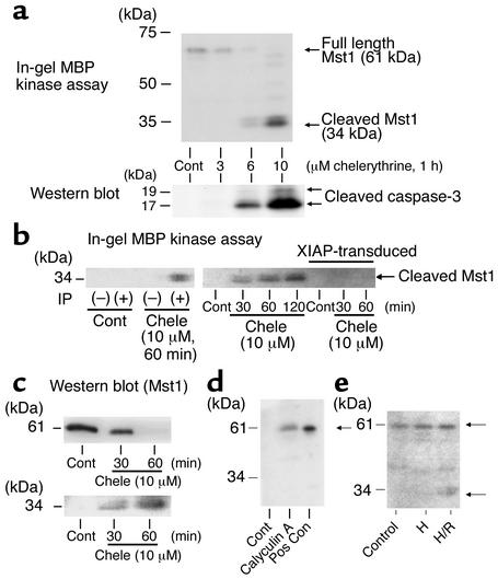
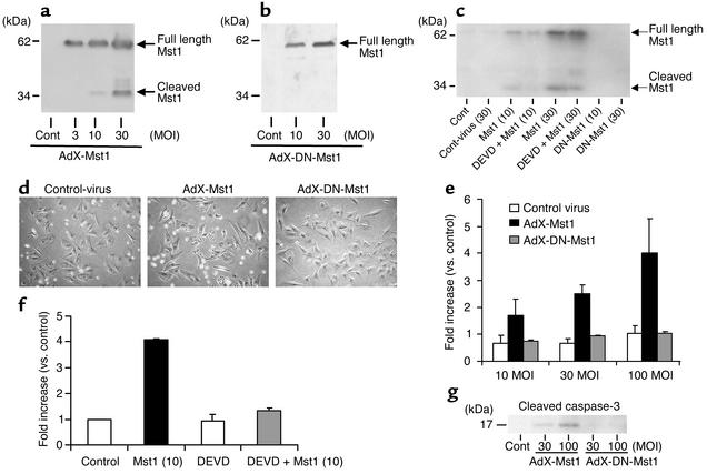
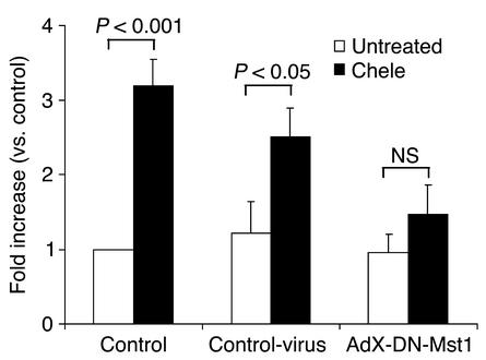
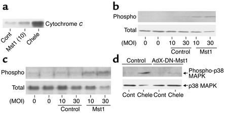
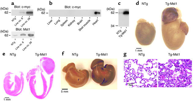
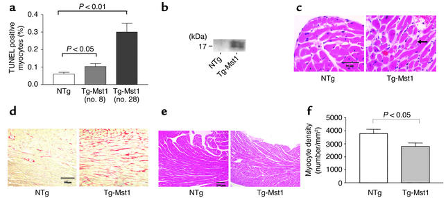
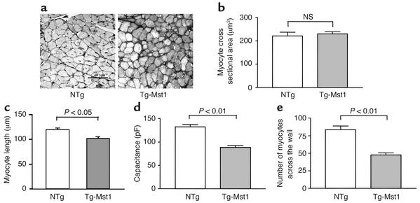
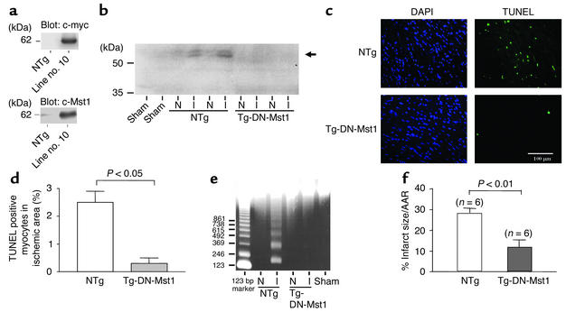
Activation of mammalian sterile 20-like kinase 1 (Mst1) by genotoxic compounds is known to stimulate apoptosis in some cell types. The importance of Mst1 in cell death caused by clinically relevant pathologic stimuli is unknown, however. In this study, we show that Mst1 is a prominent myelin basic protein kinase activated by proapoptotic stimuli in cardiac myocytes and that Mst1 causes cardiac myocyte apoptosis in vitro in a kinase activity-dependent manner. In vivo, cardiac-specific overexpression of Mst1 in transgenic mice results in activation of caspases, increased apoptosis, and dilated cardiomyopathy. Surprisingly, however, Mst1 prevents compensatory cardiac myocyte elongation or hypertrophy despite increased wall stress, thereby obscuring the use of the Frank-Starling mechanism, a fundamental mechanism by which the heart maintains cardiac output in response to increased mechanical load at the single myocyte level. Furthermore, Mst1 is activated by ischemia/reperfusion in the mouse heart in vivo. Suppression of endogenous Mst1 by cardiac-specific overexpression of dominant-negative Mst1 in transgenic mice prevents myocyte death by pathologic insults. These results show that Mst1 works as both an essential initiator of apoptosis and an inhibitor of hypertrophy in cardiac myocytes, resulting in a previously unrecognized form of cardiomyopathy.


Figures

Figure 1
Figure 1
(a) Cardiac myocytes were treated with indicated concentrations of chelerythrine for 1 h. Cell lysates were subjected to in-gel MBP kinase assay as well as immunoblotting with anti-cleaved caspase-3 Ab. Results are representative of more than five experiments. (b) Cardiac myocytes were treated with chelerythrine (Chele). In the right panel, myocytes were transduced with either control virus or adenovirus harboring XIAP 48 h before Chele application. Cell lysates were subjected to immune complex in-gel MBP kinase assays, using anti-Mst1 polyclonal Ab (pAb-15). The activity of cleaved Mst1 is shown. n = 3. (c) Cardiac myocytes were treated with Chele for the indicated durations. Immunoblot analyses were conducted using anti-Mst1 mAb (upper panel) and anti-Mst1 polyclonal Ab (lower panel), which detects the full-length form and cleaved form (amino-terminal half) of Mst1, respectively. n = 5. (d) Cardiac myocytes were treated with vehicle (lane 1), calyculin A (1 μM, lane 2), or adenovirus harboring Mst1 (10 MOI) as positive control (Pos Con, lane 3). In-gel MBP kinase assays were performed. An arrow indicates the full-length form of Mst1. n = 3. (e) Cardiac myocytes were subjected to 12 h of hypoxia alone (H) or 4 h hypoxia plus 12 h of reoxygenation (H/R). In-gel MBP kinase assays were performed. Control, Cont. Upper arrow indicates the full-length form of Mst1, while lower arrow indicates the cleaved form of Mst1. n = 3.
Figure 2
Figure 2
(ag) Cardiac myocytes were transduced with adenovirus harboring either wild-type Mst1 (AdX-Mst1), AdX-DN-Mst1, or control adenovirus (Ad5 βgal) at indicated concentrations. Myocytes were harvested 48 h after transduction. Some myocytes (c and f) were treated with a caspase-3 inhibitor (DEVD-CHO, 100 μM). (a and b) Immunoblot analyses were performed using anti-Mst1 polyclonal Ab. Cont, control, where no virus was applied. n = 4. (c) In-gel MBP kinase assays were performed. n = 5. (d) The effect of adenovirus transduction (30 MOI) upon the morphology of cardiac myocytes is shown. Note that cell death with shrinkage is observed in AdX-Mst1–transduced cardiac myocytes. n = 5. (e and f) Cytoplasmic accumulation of mono- and oligo-nucleosomes, a sensitive indicator of DNA fragmentation by apoptosis, was quantitated by Cell Death ELISA Plus. (e) Myocytes were transduced with indicated doses of either control virus, AdX-Mst1, or AdX-DN-Mst1. The experimental data are normalized by those obtained in control myocytes without adenovirus transduction. n = 3. (f) Myocytes were transduced with AdX-Mst1 (10 MOI) in the presence or absence of DEVD-CHO (100 μM). n = 3. (g) Activation of caspase-3 was determined by immunoblot analyses with anti-cleaved caspase-3 Ab. n = 3.
Figure 3
Figure 3
The effect of dominant-negative Mst1 upon chelerythrine-induced cardiac myocyte apoptosis. Cardiac myocytes were transduced with either no virus (control), control virus, or adenovirus harboring dominant-negative Mst1 (AdX-DN-Mst1). Forty-eight hours after transduction, cardiac myocytes were treated with chelerythrine (10 μM) for 1 h and cytoplasmic accumulation of mono- and oligo-nucleosome was quantitated. n = 3.
Figure 4
Figure 4
(a) Cardiac myocytes were transduced with either control virus (Cont) or AdX-Mst1 (Mst1) at 10 MOI for 48 h. As positive control, myocytes were treated with chelerythrine (Chele, 10 μM) for 1 h. The mitochondria-free cytosolic fraction was obtained. Western blot analysis was performed using anti–cytochrome c Ab as described (35). Cytochrome c oxidase IV immunoreactivity was negligible in these samples. Chele caused a release of cytochrome c to the cytosolic fraction. Expression of Mst1 increased release of cytochrome c. n = 3. (b and c) Cardiac myocytes were transduced with either control virus or AdX-Mst1(Mst1) at indicated MOIs for 48 h. Immunoblot analyses were conducted using anti-phospho p38-MAPK Ab (upper) (b) or anti-phospho JNK Ab (c). In c, phosphorylation of p46-JNK is shown. The filters were reprobed with anti–p38-MAPK Ab (b) or anti-JNK1 Ab (lower) (c). In b and c, Similar results were obtained in four experiments. (d) Cardiac myocytes were transduced with either control virus or AdX-DN-Mst1 virus. Myocytes were then stimulated with or without chelerythrine (Chele, 10 μM) for 60 min. Myocyte lysates were subjected to immunoblot analysis using anti-phospho p38-MAPK Ab. The filter was reprobed with anti–p38-MAPK Ab. Similar results were obtained in three experiments.
Figure 5
Figure 5
(a) Immunoblot analyses of heart homogenates with anti-myc Ab (upper panel). Note that Myc-Mst1 migrates at 62 kDa. Immunoblots were also conducted using anti-human Mst1 Ab (lower panel). (b) Tissue homogenates were prepared from various organs of Tg-Mst1. Immunoblot analyses were performed with anti-myc Ab. (c) Heart homogenates were prepared from Tg-Mst1 or nontransgenic control mice (NTg). In-gel MBP kinase assays were performed. n = 3. (d and e) Gross appearance and a transverse section of the hearts obtained from Tg-Mst1 and NTg. In e, hematoxylin-eosin staining was performed (96 days). (f) A photograph of the liver isolated from Tg-Mst1 and littermate NTg (96 days). (g) Hematoxylin-eosin staining of the lung.
Figure 6
Figure 6
(a) TUNEL-positive myocytes in the LV myocardium. We analyzed five Tg-Mst1 (no. 8), four Tg-Mst1 (no. 28), and nine control nontransgenic littermates (NTg). The number of TUNEL-positive myocytes was expressed as percentage of total nuclei determined by DAPI staining. (b) Heart homogenates were prepared from Tg-Mst1 and NTg. Immunoblot analyses were performed using anti-cleaved caspase-3 Ab. n = 3. (c) Hemotoxylin-eosin staining of the myocardium. Myocytes with vacuoles (shown by an arrow) are very occasionally found in the LV myocardium of Tg-Mst1. In this area, mild increases in infiltration of inflammatory cells are found. (d) Picric acid Sirius red staining of heart sections obtained from Tg-Mst1 and NTg (96 days old). (e) Hematoxylin-eosin staining of the LV myocardium. Note that infiltration of inflammatory cells are generally not significant in Tg-Mst1. (f) The number of myocytes per 1 mm2 was determined histologically from the sections obtained from LV myocardium. We studied five NTg and seven Tg-Mst1.
Figure 7
Figure 7
(a and b) LV cardiac myocyte cross sectional area was obtained from Tg-Mst1 and nontransgenic control mice (NTg) as described in Methods. Representative silver staining of the LV myocardium obtained from NTg and Tg-Mst1 are shown in a. Seven Tg-Mst1 and four NTg mice were used for the analysis in b. (c) The longitudinal myocyte length was significantly reduced in LV myocytes from Tg-Mst1 (n = 4, 111 cells) compared with NTg (n = 4, 129 cells). (d) Average cell capacitance of LV cardiac myocytes obtained from NTg and Tg-Mst1 (n = 21). (e) The number of cardiac myocyte across the LV free wall was significantly reduced in Tg-Mst1 compared with that in NTg. We studied five NTg and seven Tg-Mst1.
Figure 8
Figure 8
(a) Immunoblot analyses of heart homogenates with anti-myc Ab (upper panel) and anti-Mst1 Ab (lower panel). (bf) Tg-DN-Mst1 or NTg control mice were subjected to 20 min ischemia and 24 h of reperfusion or sham operation. (b) The heart homogenates (100 μg) obtained from ischemic (I) and nonischemic (N) areas of the LV of the mice received I/R, or from intact LV of the sham-operated mice were subjected to in-gel MBP kinase assays. I/R increased kinase activities of full-length Mst1 in the ischemic area of NTg mice, while activation of Mst1 by I/R was completely abolished in Tg-DN-Mst1. A faint band seen just above 61 kDa Mst1 most likely represents Mst2, a homologue of Mst1, whose activities are also abolished in the presence of DN-Mst1. n = 3. (c) LV tissue sections were subjected to TUNEL staining and DAPI staining. (d) TUNEL-positive myocytes in the ischemic area. The number of TUNEL-positive myocytes was expressed as percentage of total nuclei determined by DAPI staining. n = 5. (e) Genomic DNA was isolated from nonischemic (N) and ischemic (I) areas, and DNA-laddering assays were performed. The extent of DNA laddering in response to I/R was significantly smaller in Tg-DN-Mst1 compared with that in NTg. n = 3. (f) The effect of I/R upon the extent of LV myocardial infarction in Tg-DN-Mst1 and NTg. The myocardial infarction area/AAR (% infarct size/AAR) was determined as described in Methods. Note that % infarct size/AAR was significantly smaller in Tg-DN-Mst1 than in NTg.

Comment in

References

    1. Kajstura J, et al. Apoptotic and necrotic myocyte cell death are independent contributing variables of infarct size in rats. Lab. Invest. 1996;74:86–107. - PubMed
    1. Saraste A, et al. Apoptosis in human acute myocardial infarction. Circulation. 1997;95:320–323. - PubMed
    1. Tanaka M, et al. Hypoxia induces apoptosis with enhanced expression of fas antigen messenger RNA in cultured neonatal rat cardiomyocytes. Circ. Res. 1994;75:426–433. - PubMed
    1. Bialik S, et al. Myocyte apoptosis during acute myocardial infarction in the mouse localizes to hypoxic regions but occurs independently of p53. J. Clin. Invest. 1997;100:1363–1372. - PMC - PubMed
    1. Gottlieb RA, Burleson KO, Kloner RA, Babior BM, Engler RL. Reperfusion injury induces apoptosis in rabbit cardiomyocytes. J. Clin. Invest. 1994;94:1621–1628. - PMC - PubMed

Publication types

MeSH terms