. 2003 May;111(10):1497-504.
doi: 10.1172/JCI17664.

A mechanistic role for cardiac myocyte apoptosis in heart failure

Affiliations

A mechanistic role for cardiac myocyte apoptosis in heart failure

Detlef Wencker et al. J Clin Invest. 2003 May.

Abstract

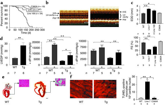
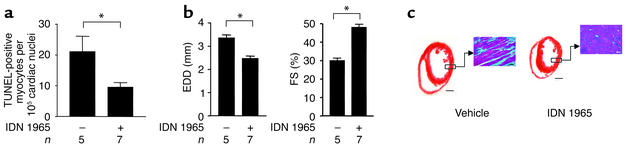
Heart failure is a common, lethal condition whose pathogenesis is poorly understood. Recent studies have identified low levels of myocyte apoptosis (80-250 myocytes per 10(5) nuclei) in failing human hearts. It remains unclear, however, whether this cell death is a coincidental finding, a protective process, or a causal component in pathogenesis. Using transgenic mice that express a conditionally active caspase exclusively in the myocardium, we demonstrate that very low levels of myocyte apoptosis (23 myocytes per 10(5) nuclei, compared with 1.5 myocytes per 10(5) nuclei in controls) are sufficient to cause a lethal, dilated cardiomyopathy. Interestingly, these levels are four- to tenfold lower than those observed in failing human hearts. Conversely, inhibition of cardiac myocyte death in this murine model largely prevents the development of cardiac dilation and contractile dysfunction, the hallmarks of heart failure. To our knowledge, these data provide the first direct evidence that myocyte apoptosis may be a causal mechanism of heart failure, and they suggest that inhibition of this cell death process may constitute the basis for novel therapies.


Figures

Figure 1
Figure 1
Expression of a conditionally active caspase-8 allele in the hearts of transgenic mice. (a) Structure of the transgene protein and strategy for its activation. M, myristoylation signal; FKBP, human FKBP-12 (pk mutant); p20 and p10, 20- and 10-kDa domains, respectively, of human procaspase-8; HA, hemagglutinin tag; FK1012, dimerizer (see text). Cysteine 360, a residue essential for caspase activity, is shown. (b) Southern blot analysis of EcoRI-digested genomic DNA from WT mice and two of the ten transgenic lines generated. The probe, an EcoRI-XbaI mouse α-cardiac myosin heavy chain genomic fragment, identified a 3.6-kb transgene fragment and a 2.5-kb fragment of the endogenous α-cardiac myosin heavy chain. (c) Immunoblot analysis using an antibody against human caspase-8, showing transgene protein expression in the hearts of the most highly expressing (line 7) and least highly expressing (line 169) lines at 3 weeks of age. Levels of the point-mutated transgene protein in the hearts of line C360A mice are similar to those of the catalytically active transgene protein in the hearts of line 7 mice. The transgene protein was not detectable in a survey of other organs, as expected with the cardiac myocyte–specific α-cardiac myosin heavy chain promoter (not shown). The lower portion of the blot was reacted with an antibody against mouse tubulin as a loading control.
Figure 2
Figure 2
Activation of the caspase transgene in vivo results in massive cardiac myocyte apoptosis and death of the animals. (a) Mortality following intraperitoneal administration of vehicle alone (81.9% polyethylene glycol 400, 9.1% Tween-80, 9% dimethylacetate) or FK1012H2 (30 mg/kg in the vehicle) to 3-week-old WT mice and transgenic line 7, 169, and C360A mice. Animals were observed for death for 7 days following FK1012H2 administration. (b) Inverse dose-dependence of the time to death following intraperitoneal administration of the indicated doses of FK1012H2 to WT or transgenic line 7 (TG) mice. Animals were observed for death for 7 days following FK1012H2 administration, with no deaths occurring in the WT group. (c) Caspase inhibition delays time to death following transgene activation. Vehicle (0.9% saline) or the polycaspase inhibitor IDN 1965 (12 mg/kg in vehicle) was administered intraperitoneally to line 7 transgenic mice 45 minutes before FK1012H2 (30 mg/kg intraperitoneally) and every 4 hours thereafter as indicated by the triangles. Bars represent the survival times of individual animals that received vehicle or IDN 1965. Animals were observed for death until all had died. *P < 0.0001. (d) Activation of the transgene caspase by FK1012H2. Immunoblot of cardiac homogenates from WT and transgenic line 7, 169, and C360A mice treated 1.5 hours earlier with vehicle or FK1012H2 (30 mg/kg intraperitoneally). Processing of procaspase-8 is indicated by disappearance of the uncleaved moiety; cleavage fragments are not reliably detected in tissue homogenates, presumably because of rapid degradation. The lower portion of the blot was reacted with an antibody against mouse tubulin as a loading control. (e) Induction of cardiac apoptosis by FK1012H2. Genomic DNA from the hearts of WT and transgenic line 7 mice 1 hour after the administration of vehicle or FK1012H2 (30 mg/kg i.v.) was size-fractionated on an agarose gel containing ethidium bromide. (f) TUNEL analysis of FK1012H2-induced apoptosis. Paraffin-embedded sections of hearts from WT and line 7 transgenic mice 1 hour after administration of vehicle or FK1012H2 (30 mg/kg i.v.). TUNEL-positive cells were primarily myocytes, but additional unidentified cells were also present that may represent degenerating myocytes or infiltrating inflammatory cells due to magnitude and rapidity of the death. Bar, 20 μM.
Figure 3
Figure 3
Very low levels of myocyte apoptosis are sufficient to cause a lethal, dilated cardiomyopathy. (a) Kaplan-Meier survival curve of WT mice, and transgenic line 7, 169, and C360A mice that have never been treated with FK1012H2. P < 0.0001 for line 7 vs. WT, line C360A, or line 169. (b) Representative two-dimensionally directed M-mode echocardiograms through the interventricular septum (IVS) and left ventricular posterior wall (PW) from 9-week-old WT and transgenic line 7 mice in the absence of FK1012H2. The electrocardiogram is shown at the bottom of each echocardiogram. (c) Quantitation of M-mode echocardiographic parameters in conscious WT and transgenic line 7, 169, and C360A mice in the absence of FK1012H2. EDD, left ventricular end-diastolic dimension; FS, fractional shortening. *P < 0.01, **P < 0.001. (d) Left ventricular hemodynamics by cardiac catheterization in 9-week-old WT and transgenic line 7 mice under basal conditions or in response to isoproterenol (500 pg i.v.), in the absence of FK1012H2. LVEDP, left ventricular end-diastolic pressure. *P < 0.02, **P < 0.002. (e) Histological analysis of 9-week-old WT and transgenic line 7 mouse hearts in the absence of FK1012H2. Coronal sections stained with H&E (bar, 1 mm), and sections from the indicated area of the left ventricular free wall stained with Masson’s trichrome (bar, 25 μm). (f) Apoptotic cardiac myocytes in WT and transgenic mice in the absence of FK1012H2. Left panels: Double staining for TUNEL (green) and desmin (red, to identify myocytes) in paraffin sections from the hearts of 9-week-old WT and line 7 transgenic mice in the absence of FK1012H2. Bar, 10 μm. Right panel: Number of TUNEL-positive cardiac myocytes per 105 nuclei in 9-week-old WT and transgenic line 7 and C360A mice in the absence of FK1012H2. *P < 0.002, **P < 0.0003.
Figure 4
Figure 4
Abrogation of dilated cardiomyopathy by caspase inhibition. Vehicle or the polycaspase inhibitor IDN 1965 (12.5 μg/h) was administered to line 7 transgenic mice by continuous subcutaneous infusion using osmotic minipumps (model 1002; ALZET Corp., Cupertino, California, USA), beginning at 3.5–4.0 weeks of age, when cardiac dimensions, function, and histology are normal, and continuing until sacrifice at 7.5–8.0 weeks of age, when these transgenic mice uniformly exhibit a severe dilated cardiomyopathy. At 7.5–8.0 weeks of age, echocardiography, TUNEL, and histological examination of cardiac tissue were performed. (a) Number of TUNEL-positive cardiac myocytes per 105 nuclei in vehicle- and IDN 1965–treated line 7 mice. *P < 0.03. (b) M-mode echocardiographic parameters from vehicle- and IDN 1965–treated line 7 mice. *P < 0.0003. (c) Coronal sections from vehicle- and IDN 1965–treated line 7 mice stained with H&E (bar, 1 mm), and sections from the indicated area of the left ventricular free wall stained with Masson’s trichrome (bar, 20 μm).

Comment in

References

    1. Braunwald E, Bristow MR. Congestive heart failure: fifty years of progress. Circulation. 2000;102:IV14–IV23. - PubMed
    1. Chien KR. Stress pathways and heart failure. Cell. 1999;98:555–558. - PubMed
    1. Lefkowitz RJ, Rockman HA, Koch WJ. Catecholamines, cardiac beta-adrenergic receptors, and heart failure. Circulation. 2000;101:1634–1637. - PubMed
    1. Marks AR. Ryanodine receptors, FKBP12, and heart failure. Front. Biosci. 2002;7:d970–d977. - PubMed
    1. Luo W, et al. Targeted ablation of the phospholamban gene is associated with markedly enhanced myocardial contractility and loss of beta-agonist stimulation. Circ. Res. 1994;75:401–409. - PubMed

Publication types

MeSH terms