. 2003 May;111(10):1505-18.
doi: 10.1172/JCI17662.

CD137 costimulatory T cell receptor engagement reverses acute disease in lupus-prone NZB x NZW F1 mice

Affiliations

CD137 costimulatory T cell receptor engagement reverses acute disease in lupus-prone NZB x NZW F1 mice

Juergen Foell et al. J Clin Invest. 2003 May.

Abstract

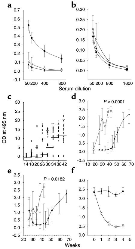
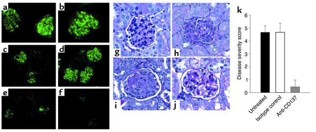
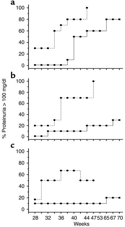
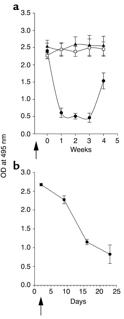
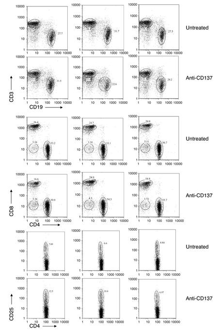
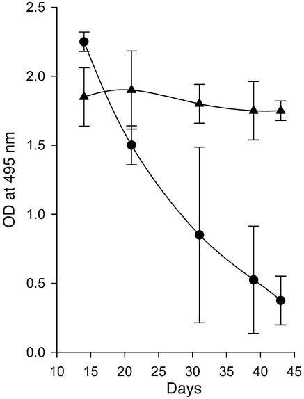
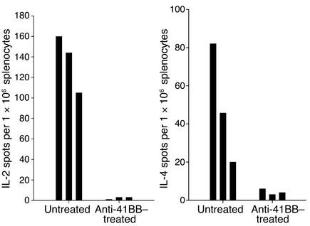
Systemic lupus erythematosus (SLE) is a CD4(+) T cell-dependent, immune complex-mediated, autoimmune disease that primarily affects women of childbearing age. Generation of high-titer affinity-matured IgG autoantibodies, specific for double-stranded DNA and other nuclear antigens, coincides with disease progression. Current forms of treatment of SLE including glucocorticosteroids are often inadequate and induce severe side effects. Immunological approaches for treating SLE in mice using anti-CD4 mAb's or CTLA4-Ig and anti-CD154 mAb's have proven to be effective. However, like steroid treatment, these regimens induce global immunosuppression, and their withdrawal allows for disease progression. In this report we show that lupus-prone NZB x NZW F(1) mice given three injections of anti-CD137 (4-1BB) mAb's between 26 and 35 weeks of age reversed acute disease, blocked chronic disease, and extended the mice's lifespan from 10 months to more than 2 years. Autoantibody production in recipients was rapidly suppressed without inducing immunosuppression. Successful treatment could be traced to the fact that NZB x NZW F(1) mice, regardless of their age or disease status, could not maintain pathogenic IgG autoantibody production in the absence of continuous CD4(+) T cell help. Our data support the hypothesis that CD137-mediated signaling anergized CD4(+) T cells during priming at the DC interface.


Figures

Figure 1
Figure 1
Inhibition of autoantibody production by anti-CD137 mAb’s. NZB/W F1 mice were injected intraperitoneally with anti-CD137 or control mAb’s, and serum samples were collected and assayed in triplicate by ELISA for anti-dsDNA antibodies. IgG or IgM anti-dsDNA autoantibody titers are expressed as the mean ± SD measuring the OD, at 495 nm, of triplicates for individual mice, using serially diluted serum samples. (a) Anti-dsDNA IgG serum titers of 41-week-old NZB/W F1 mice that were injected at weeks 14, 17, 20, 23, and 26 with anti-CD137 (open squares) or isotype-matched control mAb (filled circles), compared with serum from BALB/c mice (filled triangles). (b) Kinetics of IgM production in anti-CD137 mAb–treated mice (filled circles), mice treated with isotype-matched control mAb’s (open squares), and PBS-treated mice (filled triangles). (c) Kinetics of the appearance of IgG dsDNA-reactive autoantibodies in NZB/W F1 mice that received a single 200-μg intraperitoneal injection of anti-CD137 (filled squares) or control mAb (open squares) at 8 weeks of age. (d) Kinetics of IgG anti-dsDNA autoantibody production in mice in which anti-CD137 treatment (filled triangles) or isotype control mAb (open circles) 8-week-old were treated through 25 weeks of age (200 μg injected intraperitoneally every third week). (e) Kinetics of anti-dsDNA autoantibody production anti-CD137 treated (filled triangles) or control (open circles) mAb treated at 26 weeks of age after they had developed autoantibodies in their serum. (f) Clearance of serum anti-dsDNA IgG autoantibodies in proteinuric 40-week-old NZB/W F1 mice treated with a single injection of anti-CD137 (open triangles) or control mAb’s (filled circles).
Figure 2
Figure 2
(af) Anti-CD137 mAb’s block immune complex formation. Kidneys sections from NZB/W F1 mice at 35 (a, c, and e) and 45 (b, d, and f) weeks of age that had received no treatment (a and b), isotype-matched control mAb’s (c and d), or anti-CD137 mAb’s (e and f) were stained with FITC-goat anti-mouse IgG. (gj) Anti-CD137 mAb’s block kidney disease. Shown are glomeruli from a BALB/c mouse (g), an untreated NZB/W F1 mouse (h), an isotype control-treated mouse (i), and an anti-CD137 mAb–treated mouse (j). (k) Disease severity following anti-CD137 mAb treatment. NZB/W F1 mice were untreated or treated with either isotype-matched control mAb’s or anti-CD137 mAb’s beginning at 14 weeks of age. Disease index was based upon kidney histopathology (see Methods).
Figure 3
Figure 3
Anti-CD137 mAb treatment blocks germinal center reactions. NZB/W F1 mice were injected once every third week with anti-CD137 or isotype-matched control mAb’s beginning at 14 weeks of age and continuing through 26 weeks of age. The mice were euthanized, and spleens were removed for thin sectioning and H&E staining. Sections were viewed with ×20 and ×40 objectives and photographed. (a and c) Representative fields at ×20 and ×40, respectively, of mice injected with isotype-matched control mAb’s. (b and d) Representative fields at ×20 and ×40, respectively, of mice treated with anti-CD137 mAb’s. Bar, 100 μg.
Figure 4
Figure 4
Anti-CD137 mAb treatment protects mice from developing proteinuria. Urine was collected from mice in each of the three groups enrolled in the serum anti-dsDNA autoantibody studies described in Figure 1 and tested for protein content by the Albustix method as described in Methods. (ac) Ovals represent untreated mice and squares represent mice treated with anti-CD137 mAb’s. (a) One injection of anti-CD137 mAb was given at 8 weeks of age. (b) Mice received seven injections of anti-CD137 mAb’s beginning at 8 weeks of age and continuing once every third week. (c) Mice received their first of three anti-CD137 mAb injections at 25 weeks of age and one thereafter every third week for a total of three injections. All injections were administered intraperitoneally at a dose of 200 μg.
Figure 5
Figure 5
(a) CD4+ T cell depletion decreases serum anti-dsDNA autoantibodies. Thirty-six- to 40-week-old NZB/W F1 mice with high titers of anti-dsDNA antibodies were depleted of CD4+ T cells or CD8+ T cells by four intraperitoneal injections of anti-CD4 mAb (filled circles) or anti-CD8 mAb (open squares) every 3 days, or left untreated (filled triangles). After the last injection of antibody (arrow, x axis), serum samples were collected as indicated and assayed in triplicate for anti-dsDNA reactive antibodies. IgG anti-dsDNA autoantibody titers as a function of time and treatment are expressed as the mean ± SD measured at an OD at 495 nm of serum from individual mice, using a 1:100 diluted serum sample. (b) Anti-CD137 mAb’s suppress anti-dsDNA production in the absence of CD8+ T cells. CD8+ T cells were depleted from NZB/W F1 mice prior to treatment with anti-CD137 mAb’s to determine whether CD8+ regulatory T cells were generated in response to CD137-mediated T cell costimulation. Mice depleted of CD8+ T cells as described above were injected with 200 μg anti-CD137 mAb and assayed for anti-dsDNA autoantibodies in their serum over a 23-day period.
Figure 6
Figure 6
Anti-CD137 mAb’s do not delete or alter the frequency of T or B cells. Six-month-old NZB/W F1 mice with prominent signs of proteinuria were injected intraperitoneally with 200 μg anti-CD137 mAb’s or left untreated. Three days or 21 days later (only day 3 data are shown, as there were no differences between the two data sets), the mice were euthanized, and spleen-derived lymphocytes were phenotyped using a dual-laser multiparameter FACSCalibur analytical flow cytometer (Becton Dickinson Immunocytometry Systems, San Jose, California, USA) for single-cell expression of the following cell surface receptors: CD3, CD4, CD8, CD25, and CD19.
Figure 7
Figure 7
Anti-CD137 mAb’s do not suppress established anti-SRBC humoral immune responses. Five 26-week-old NZB/W F1 mice were immunized with SRBC and boosted at 28 weeks of age. At 30 weeks of age, the mice were injected with 200 μg anti-CD137 mAb’s intraperitoneally. Serum from the mice was collected at various time points after treatment and assayed by ELISA for anti-SRBC (triangles) or anti-dsDNA (circles) antibodies.
Figure 8
Figure 8
Anti-CD137 mAb treatment suppresses IL-2 and IL-4 production. Twenty-week-old NZB/W F1 mice were injected intraperitoneally with 200 μg anti-CD137 or isotype-matched control mAb. The mice were euthanized 5 days later, and IL-4 and IL-2 production was measured over a 36-hour period by ELISpot assays using spleen- and lymph node–derived CD4+ T cells. Shown are the responses of individual mice treated or not treated with anti-CD137 mAb’s. The results are highly reproducible and typical of multiple experiments performed.
Figure 9
Figure 9
Survival and homing of CD4+ T cells and B cells. T and B cells were obtained from 26-week-old NZB/W F1 or BALB/c mice, labeled with CFSE, and injected i.v. into age-, sex-, and strain-matched recipients. At 72 hours, recipient mice were bled and euthanized. Single-cell suspensions of spleen, lymph node, bone marrow, and PBMCs were stained with phycoerythrin-conjugated anti-CD19 or -CD4 mAb’s, and their frequency was determined by flow cytometry. CFSE fluorescence intensity was measured, and proliferation of each cell population was determined based on the incremental loss of CFSE fluorescence. The far-left peaks in each histogram represent either dead or dying cells, which were present within 24 hours of culture; the extreme loss of fluorescence could not be attributed to cell division in so short a period of time.
Figure 10
Figure 10
Adoptive transfer of primed CD4+ T cells bypasses CD137-induced suppression. CD4+ T cells from 26-week-old NZB/W F1 untreated diseased mice were used as a source of dsDNA-primed Th cells. Age-matched mice (n = 10) that had been injected 2 weeks earlier with anti-CD137 were injected i.v. with 1 × 10⁷ T cells (filled circles). A second group of mice received only anti-CD137 mAb’s (open triangles). The mice were subsequently bled on a weekly basis, and serum anti-dsDNA levels were determined by ELISA.
Figure 11
Figure 11
Adoptive transfer of naive bone marrow–derived DCs bypasses CD137-induced suppression. One million NZB/W F1 bone marrow–derived CD11c+ DCs (15–30% CD137+) were injected i.v. into 26-week-old NZB/W F1 mice (n = 10) that had been injected with anti-CD137 mAb’s 4 weeks earlier. The mice were bled weekly, and anti-dsDNA titers were determined as above.

Similar articles

Cited by

References

    1. Wakeland EK, Liu K, Graham RR, Behrens TW. Delineating the genetic basis of systemic lupus erythematosus. Immunity. 2001;15:397–408. - PubMed
    1. Stott DI, Merino J, Schurmans S, Lambert PH. Expression of anti-DNA clonotypes and the role of helper T-lymphocytes during the autoimmune response in mice tolerant to alloantigens. Autoimmunity. 1988;1:253–266. - PubMed
    1. Channing AA, Kasuga T, Horowitz RE, Dubois EL, Demopoulos HB. An ultrastructural study of spontaneous lupus nephritis in the NZB-BL-NZW mouse. Am. J. Pathol. 1965;47:677–694. - PMC - PubMed
    1. Humbert M, Galanaud P. B-lymphocyte hyperreactivity and differentiation factors of T-lymphocytes in systemic lupus erythematosus. Ann. Med. Interne (Paris). 1990;141:213–216. - PubMed
    1. Sobel ES, et al. T-B collaboration for autoantibody production in lpr mice is cognate and MHC-restricted. J. Immunol. 1994;152:6011–6016. - PubMed

Publication types

MeSH terms