Case Reports
. 2003 May;111(10):1537-45.
doi: 10.1172/JCI16387.

Pacemaker channel dysfunction in a patient with sinus node disease

Affiliations
Case Reports

Pacemaker channel dysfunction in a patient with sinus node disease

Eric Schulze-Bahr et al. J Clin Invest. 2003 May.

Abstract

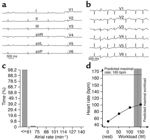
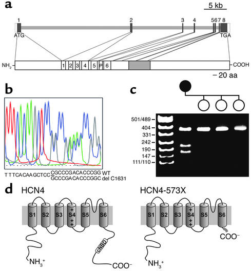
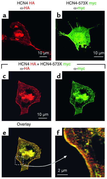
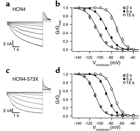
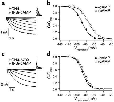
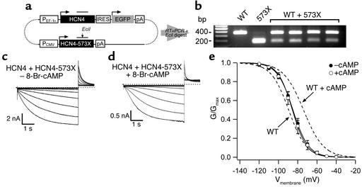
The cardiac pacemaker current I(f) is a major determinant of diastolic depolarization in sinus nodal cells and has a key role in heartbeat generation. Therefore, we hypothesized that some forms of "idiopathic" sinus node dysfunction (SND) are related to inherited dysfunctions of cardiac pacemaker ion channels. In a candidate gene approach, a heterozygous 1-bp deletion (1631delC) in exon 5 of the human HCN4 gene was detected in a patient with idiopathic SND. The mutant HCN4 protein (HCN4-573X) had a truncated C-terminus and lacked the cyclic nucleotide-binding domain. COS-7 cells transiently transfected with HCN4-573X cDNA indicated normal intracellular trafficking and membrane integration of HCN4-573X subunits. Patch-clamp experiments showed that HCN4-573X channels mediated I(f)-like currents that were insensitive to increased cellular cAMP levels. Coexpression experiments showed a dominant-negative effect of HCN4-573X subunits on wild-type subunits. These data indicate that the cardiac I(f) channels are functionally expressed but with altered biophysical properties. Taken together, the clinical, genetic, and in vitro data provide a likely explanation for the patient's sinus bradycardia and the chronotropic incompetence.


Figures

Figure 1
Figure 1
(a) Baseline electrocardiogram of the index patient showing marked sinus bradycardia (41 bpm). (b) ECG recorded during an episode of intermittent atrial fibrillation. (c) P-wave histograms obtained from pacemaker interrogation after implantation. At day 68, 98% of the atrial rates were below a threshold level of 60 bpm. (d) Heart rates (at sinus rhythm) of the index patient during bicycle exercise, before pacemaker implantation. The vertical dotted line and gray area indicate the predicted mean maximal work performance ± SD (with respect to the patient’s age [65–70 years] and gender) (25). The dotted horizontal line indicates the predicted heart rate value during maximal work load for women 66 years of age (25).
Figure 2
Figure 2
Mutation detection in the HCN4 gene. (a) The human HCN4 gene consists of eight exons. The minimal exon size was 141 bp (exon 5), and the maximal size was 1,465 bp (exon 8); the largest intron was intron 1 (∼24 kb) and the smallest intron 5 (102 bp). The functional domains of the wild-type HCN4 channel are delineated below. P, pore region; 1–6, transmembrane domains. (b) Electropherogram after direct sequencing of an index patient with SND. A heterozygous 1-bp deletion (1631delC) in HCN4 resulted in a superimposing sequence pattern consisting of the wild-type and mutant exon 5 sequence. (c) The heterozygous deletion mutation induces an EciI restriction site in exon 5; after restriction enzyme analysis, the uncut wild-type fragment (380 bp) and two EciI fragments (200 and 180 bp, cut mutant fragment) were found in the index patient. Her three healthy children had the wild-type configuration. The left lane shows the size standard in base pairs (pUC19 DNA/MspI). (d) Schematic topology of HCN4 channels with six transmembrane segments (S1–S6) and intracellular N- and C-termini. Because of the reading frame shift in the nucleotide sequence, a resulting premature stop codon deleted the C-terminally located cNBD in HCN4-573X that is replaced by 29 novel amino acids (thick gray line). The relative sizes of the transmembrane segments and N- or C-termini are not drawn to scale.
Figure 3
Figure 3
Immunostaining of HCN4 and HCN4-573X in COS-7 cells. Wild-type and HCN4-573X channels were transiently expressed in COS-7 cells. For detection, wild-type HCN4 was HA-tagged and HCN4-573X was myc-tagged. Both channels showed comparable immunostains (a and b) with immunoreactivity detectable in perinuclear regions and at the plasma membrane. When coexpressed (cf), the immunostaining patterns did not change and completely overlapped, and they were particularly concentrated in the cell membrane (f).
Figure 4
Figure 4
Functional characterization of homomeric HCN4 and HCN4-573X channels in COS-7 cells under control conditions. Representative current traces of wild-type (a) or HCN4-573X (c) channels elicited by 5-second-long hyperpolarizing voltage steps from –40 mV to –120 mV under control conditions. Relative open probability of wild-type (b) and HCN4-573X (d) channels was dependent on test-pulse duration (gray circles, 2 seconds; black circles, 5 seconds; white circles, 15 seconds). Absence of error bars indicates errors smaller than the symbol size.
Figure 5
Figure 5
Functional characterization of homomeric wild-type and HCN4–573X channels in COS-7 cells in the presence of 8-Br-cAMP. Shown are representative current traces of wild-type (a) or HCN4-573X (c) channels elicited by 5-second-long hyperpolarizing voltage steps from –40 mV to –120 mV in the presence of 8-Br-cAMP. Voltage dependence of wild-type (b) and HCN4-573X (d) conductances in the absence (black circles) or presence (white circles) of 1 mM 8-Br-cAMP. Absence of error bars indicates errors smaller than the symbol size.
Figure 6
Figure 6
Functional characterization of coexpressed HCN4 and HCN4-573X subunits in COS-7 cells. (a) Schematic representation of the expression construct used for coexpression experiments (not drawn to scale). Wild-type and mutant HCN4 cDNAs are located on the same plasmid (indicated with dotted lines); initiation codons of open reading frames are indicated with horizontal arrows. Structural elements such as promoters (PCMV, PEF-1α), IRES, and polyadenylation signals are shown in open boxes. The PCR products amplified from reverse-transcribed total RNA are indicated by a horizontal line above the HCN4 open reading frames. The location of the EciI restriction site is indicated by a vertical arrow. pA, polyadenylation signal. (b) Agarose gel electrophoresis of EciI-digested RT-PCR products amplified from total RNA of transiently transfected COS-7 cells. The wild-type fragment has a size of 353 bp and contains no EciI restriction site. In contrast, the 352-bp-long mutant PCR fragment (573X) is digested by EciI (see Figure 2c). The resulting two fragments (160 bp and 192 bp) are not separated under the chosen conditions. Shown are representative current traces of coexpressed wild-type and HCN4-573X subunits elicited by hyperpolarizing voltage steps from –40 mV to –120 mV in the absence (c) or presence (d) of 8-Br-cAMP. Voltage dependences of heteromeric wild-type/hHCN4-573X (e) conductances in the absence (black circles) or presence (white circles) of 1 mM 8-Br-cAMP are shown. The dashed lines indicate the respective Boltzmann fits of HCN4 (WT) with or without application of 8-Br-cAMP taken from Figure 4. Absence of error bars indicates errors smaller than the symbol size.

References

    1. Lamas GA, et al. The mode selection trial (MOST) in sinus node dysfunction: design, rationale, and baseline characteristics of the first 1000 patients. Am. Heart J. 2000;140:541–551. - PubMed
    1. Sarachek NS, Leonard JL. Familial heart block and sinus bradycardia. Classification and natural history. Am. J. Cardiol. 1972;29:451–458. - PubMed
    1. Spellberg RD. Familial sinus node disease. Chest. 1971;60:246–251. - PubMed
    1. Lehmann H, Klein UE. Familial sinus node dysfunction with autosomal dominant inheritance. Br. Heart J. 1978;40:1314–1316. - PMC - PubMed
    1. Mackintosh AF, Chamberlain DA. Sinus node disease affecting both parents and both children. Eur. J. Cardiol. 1979;10:117–122. - PubMed

Publication types

MeSH terms