

D-serine and serine racemase are present in the vertebrate retina and contribute to the physiological activation of NMDA receptors
- PMID: 12750462
- PMCID: PMC164525
- DOI: 10.1073/pnas.1237052100
D-serine and serine racemase are present in the vertebrate retina and contribute to the physiological activation of NMDA receptors
Abstract
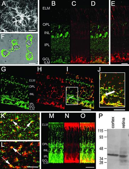
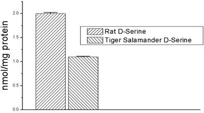
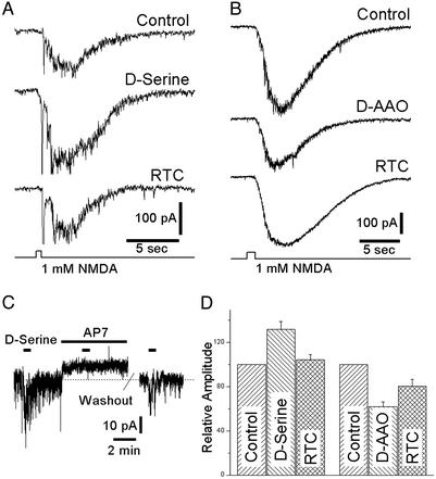
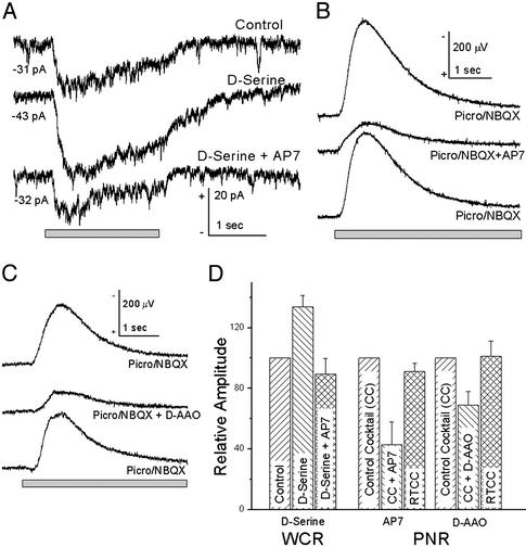
d-serine has been proposed as an endogenous modulator of N-methyl-d-aspartate (NMDA) receptors in many brain regions, but its presence and function in the vertebrate retina have not been characterized. We have detected d-serine and its synthesizing enzyme, serine racemase, in the retinas of several vertebrate species, including salamanders, rats, and mice and have localized both constituents to Müller cells and astrocytes, the two major glial cell types in the retina. Physiological studies in rats and salamanders demonstrated that, in retinal ganglion cells, d-serine can enhance excitatory currents elicited by the application of NMDA, as well as the NMDA receptor component of light-evoked synaptic responses. Application of d-amino acid oxidase, which degrades d-serine, reduced the magnitude of NMDA receptor-mediated currents, raising the possibility that endogenous d-serine serves as a ligand for setting the sensitivity of NMDA receptors under physiological conditions. These observations raise exciting new questions about the role of glial cells in regulating the excitability of neurons through release of d-serine.
Figures
Similar articles
-
A new strategy to decrease N-methyl-D-aspartate (NMDA) receptor coactivation: inhibition of D-serine synthesis by converting serine racemase into an eliminase.Proc Natl Acad Sci U S A. 2001 Apr 24;98(9):5294-9. doi: 10.1073/pnas.091002298. Epub 2001 Apr 17. Proc Natl Acad Sci U S A. 2001. PMID: 11309496 Free PMC article.
-
D-serine regulates CREB phosphorylation induced by NMDA receptor activation in Müller glia from the retina.Neurosci Lett. 2007 Oct 29;427(1):55-60. doi: 10.1016/j.neulet.2007.09.009. Epub 2007 Sep 15. Neurosci Lett. 2007. PMID: 17920195
-
Retinal NMDA receptor function and expression are altered in a mouse lacking D-amino acid oxidase.J Neurophysiol. 2013 Dec;110(12):2718-26. doi: 10.1152/jn.00310.2013. Epub 2013 Sep 25. J Neurophysiol. 2013. PMID: 24068757 Free PMC article.
-
Structure-function relationships in human D-amino acid oxidase.Amino Acids. 2012 Nov;43(5):1833-50. doi: 10.1007/s00726-012-1345-4. Epub 2012 Aug 3. Amino Acids. 2012. PMID: 22865246 Review.
-
Serine racemase as a prime target for age-related memory deficits.Eur J Neurosci. 2013 Jun;37(12):1931-8. doi: 10.1111/ejn.12226. Eur J Neurosci. 2013. PMID: 23773062 Review.
Cited by
-
Promising Application of D-Amino Acids toward Clinical Therapy.Int J Mol Sci. 2022 Sep 16;23(18):10794. doi: 10.3390/ijms231810794. Int J Mol Sci. 2022. PMID: 36142706 Free PMC article. Review.
-
D-serine uptake and release in PC-12 cells measured by chiral microchip electrophoresis-mass spectrometry.ACS Chem Neurosci. 2015 Apr 15;6(4):582-7. doi: 10.1021/cn5003122. Epub 2015 Feb 3. ACS Chem Neurosci. 2015. PMID: 25611520 Free PMC article.
-
Eph signaling regulates gliotransmitter release.Commun Integr Biol. 2011 Mar;4(2):223-6. doi: 10.4161/cib.4.2.14507. Commun Integr Biol. 2011. PMID: 21655447 Free PMC article.
-
Astrocyte, the star avatar: redefined.J Biosci. 2008 Sep;33(3):405-21. doi: 10.1007/s12038-008-0060-5. J Biosci. 2008. PMID: 19005240 Review.
-
Glycine transporter 1 modulates GABA release from amacrine cells by controlling occupancy of coagonist binding site of NMDA receptors.J Neurophysiol. 2013 Sep;110(6):1393-403. doi: 10.1152/jn.00193.2013. Epub 2013 Jun 26. J Neurophysiol. 2013. PMID: 23803324 Free PMC article.
References
-
- Corrigan, J. J. (1969) Science 164, 142-149. - PubMed
-
- Hashimoto, A., Nishikawa, T., Oka, T., Takahashi, K. & Hayashi, T. (1992) J. Chromatogr. 582, 41-48. - PubMed
-
- Hashimoto, A., Nishikawa, T., Oka, T. & Takahashi, K. (1993) J. Neurochem. 60, 783-786. - PubMed
-
- Kleckner, N. W. & Dingledine, R. (1988) Science 241, 835-837. - PubMed
Publication types
MeSH terms
Substances
Grants and funding
LinkOut - more resources
Full Text Sources
Other Literature Sources
