. 2003 May 27;100(11):6837-42.
doi: 10.1073/pnas.1132113100. Epub 2003 May 15.

Conservation and molecular dissection of ROUGH SHEATH2 and ASYMMETRIC LEAVES1 function in leaf development

Affiliations

Conservation and molecular dissection of ROUGH SHEATH2 and ASYMMETRIC LEAVES1 function in leaf development

George Theodoris et al. Proc Natl Acad Sci U S A. .

Abstract

Maize ROUGH SHEATH2 (RS2) and Arabidopsis ASYMMETRIC LEAVES1 (AS1) are orthologous Myb-related genes required for leaf development and act as negative regulators of class 1 KNOTTED1-like homeobox (KNOX) genes in leaf primordia. Expression of RS2 in Arabidopsis fully complements as1 leaf phenotypes and represses the expression of the KNOX gene KNAT1 in leaves. Whereas loss of AS1 function in Arabidopsis results in rounded, lobed leaves with shorter and wider petioles, overexpression of either RS2 or AS1 results in longer and narrower leaves with longer petioles than wild type. A conserved C-terminal domain (CTD) mediates homodimerization of both RS2 and AS1 and modulates leaf shape when expressed independently of the Myb domain in Arabidopsis. Homodimerization is not absolutely required for KNAT1 repression. RS2:GFP fusion protein is biologically active, localized in discrete dynamic subnuclear foci and associates with DNA during cell division.


Figures
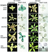
Fig. 1.
Fig. 1.
Phenotype (A, C, E, G, I, K, N, Q, T, and W) and KNAT1:GUS (B, D, F, H, J, L, N, P, R, and T) expression in as1 mutant (AJ) and wild type (KT) plants containing RS2 and AS1 transgenes. (A and B) as1 mutant; (C and D) as1 mutant + 35S:AS1;(E and F) as1 mutant + 35S:RS2;(G and H) as1 mutant + RS2ΔMYB;(I and J) as1 mutant + RS2ΔCTD; (K and L) wild type; (M and N) wild type + 35S:AS1; (O and P) wild type + 35S:RS2; (Q and R) wild type + RS2ΔMYB; (S and T) wild type + RS2ΔCTD.
Fig. 2.
Fig. 2.
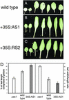
Phenotypic consequences of overexpression of AS1 and RS2. Juvenile rosette leaves of wild-type plants transformed with vector alone (A), 35S:AS1 (B), and 35S:RS2 (C). (D) Graph of leaf dimensions of wild-type, as1 mutant, and 35S:AS1 plants. The left bars represent the percentage of leaf length comprised of petiole and the right bars represent the leaf width/length ratio.
Fig. 3.
Fig. 3.
(A) Two-hybrid deletion analysis of RS2 and AS1 homodimerization. Interaction is observed only between bait and prey plasmids containing the conserved full-length RS2 or AS1 or the conserved CTD. β-Galactosidase activity is expressed as nmol of o-nitrophenyl-d-galactoside (ONPG) hydrolyzed min-1·mg-1 total protein. (B) SDS/PAGE of 35S-labeled RS2 protein under reducing and nonreducing conditions. Lane 1, RS2 after reduction with 100 mM DTT; lane 2, RS2 without treatment with a reducing agent.
Fig. 4.
Fig. 4.
Suppression of the as1 leaf phenotype by overexpression of RS2ΔCTD and RS2ΔMYB. Representative leaves from as1 mutant plants transformed with vector alone (A), 35S:RS2ΔCTD (B), and 35S:RS2ΔMYB (C) and a wild-type plant (D) are shown.
Fig. 5.
Fig. 5.
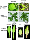
Phenotypic consequences of overexpression of RS2ΔMYB on leaf development. Shown are the first true leaves of a wild-type sib (A) and two 35S:RS2ΔMYB (B) plants. (C and D) Rosette phenotypes of older mature plants: wild type transformed with vector alone (C) and 35S:RS2ΔMYB (D). Adult leaf morphology of wild type (E), wild type + 35S:RS2ΔMYB (F), as1 (G), and as1 + 35S:RS2ΔMYB (H) is shown.
Fig. 6.
Fig. 6.
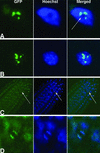
Subcellular localization of RS2:GFP. Nuclei are visualized with Hoechst stain. (A and B) Localization of RS2:GFP fusion protein in young leaf cells. The subnuclear bodies are present in the nucleoplasm (B) and around the periphery of the nucleolus, which is marked by an arrow in A.(C) Confocal image of a root tip showing a dividing cell marked by arrows. (D) A close-up showing localization of fusion protein with dividing chromosomes.

References

    1. Waites, R., Selvadurai, H. R. N., Oliver, I. R. & Hudson, A. (1998) Cell 93, 779-789. - PubMed
    1. Schneeberger, R., Tsiantis, M., Freeling, M. & Langdale, J. A. (1998) Development (Cambridge, U.K.) 125, 2857-2865. - PubMed
    1. Timmermans, M. C. P., Hudson, A., Becraft, P. W. & Nelson, T. (1999) Science 284, 151-153. - PubMed
    1. Tsiantis, M., Schneeberger, R., Golz, J. F., Freeling, M. & Langdale, J. A. (1999) Science 284, 154-156. - PubMed
    1. Byrne, M. E., Barley, R., Curtis, M., Arroyo, J. M., Dunham, M., Hudson, A. & Martienssen, R. A. (2000) Nature 408, 967-971. - PubMed