

The locus coeruleus complex of the bottlenose dolphin (Tursiops truncatus) as revealed by tyrosine hydroxylase immunohistochemistry
- PMID: 12753352
- PMCID: PMC8799625
- DOI: 10.1046/j.1365-2869.2003.00350.x
The locus coeruleus complex of the bottlenose dolphin (Tursiops truncatus) as revealed by tyrosine hydroxylase immunohistochemistry
Abstract
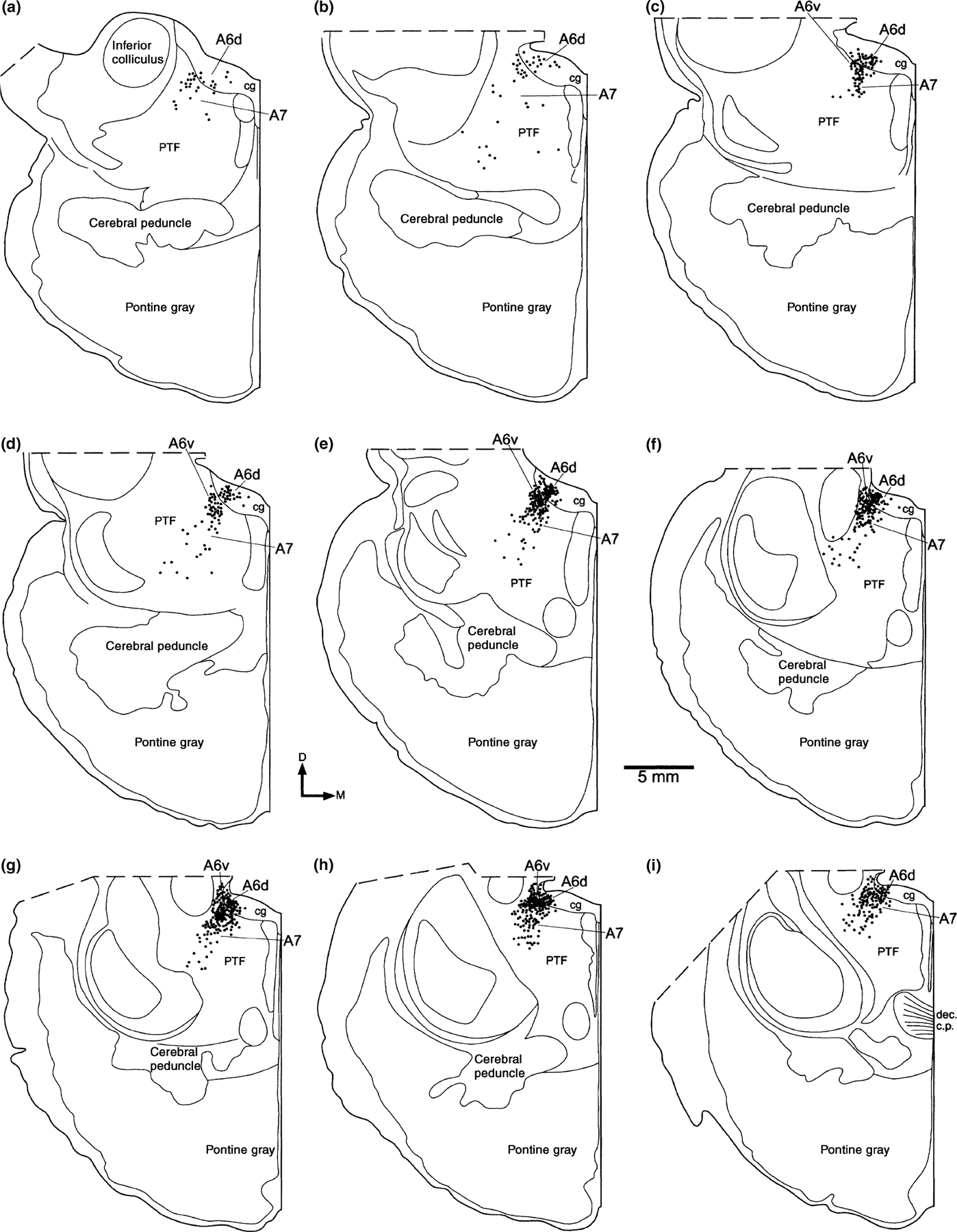
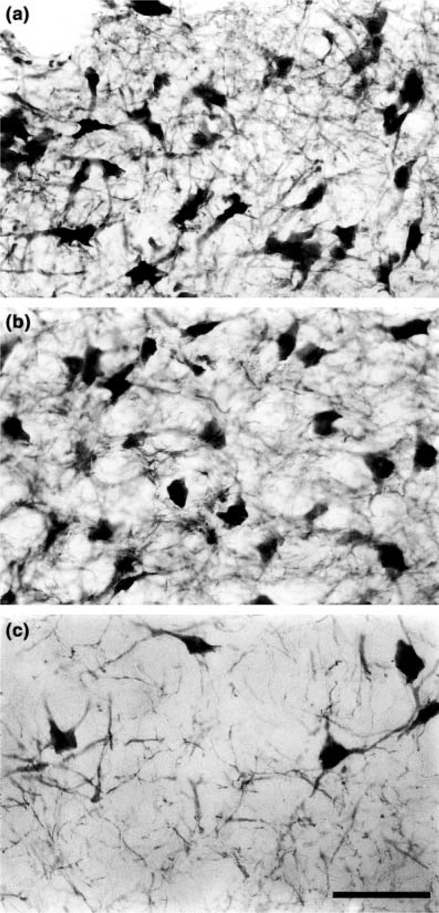
Using tyrosine hydroxylase immunohistochemistry we examined the structure of the pontine, or rostral rhombencephalic, catecholaminergic cells groups, which may be collectively termed the locus coeruleus complex (LC), in the bottlenose dolphin. The present study is the first to describe the LC in a cetacean species and, at 1.3 kg, represents the largest non-human brain to date in which the LC has been investigated. We identified four catecholaminergic cell groups in the dorsal pontine tegementum and peri-aqueductal gray matter: A6 dorsal (locus coeruleus), A6 ventral (locus coeruleus alpha), A7 (subcoeruleus), and A5 (fifth arcuate nucleus). No patterns of cellular distribution, nuclear subdivision, or cellular morphology indicate specialization of the LC, which might have been anticipated because of the large absolute brain size and unihemispheric sleep phenomenology of cetaceans.
Figures
Similar articles
-
Locus coeruleus complex of the family Delphinidae.Sci Rep. 2018 Apr 3;8(1):5486. doi: 10.1038/s41598-018-23827-z. Sci Rep. 2018. PMID: 29615733 Free PMC article.
-
Nuclear parcellation of pontine catecholaminergic and cholinergic neurons in gray parrots and pied crow brains.Anat Rec (Hoboken). 2025 Sep;308(9):2433-2449. doi: 10.1002/ar.25593. Epub 2024 Oct 23. Anat Rec (Hoboken). 2025. PMID: 39440441 Free PMC article.
-
Fos expression in pontomedullary catecholaminergic cells following rapid eye movement sleep-like episodes elicited by pontine carbachol in urethane-anesthetized rats.Neuroscience. 2008 Mar 3;152(1):208-22. doi: 10.1016/j.neuroscience.2007.11.013. Neuroscience. 2008. PMID: 18155849 Free PMC article.
-
The distribution and morphological characteristics of catecholaminergic cells in the diencephalon and midbrain of the bottlenose dolphin (Tursiops truncatus).Brain Behav Evol. 2004;64(1):42-60. doi: 10.1159/000077542. Epub 2004 Mar 26. Brain Behav Evol. 2004. PMID: 15051966 Free PMC article.
-
The Mammalian Locus Coeruleus Complex-Consistencies and Variances in Nuclear Organization.Brain Sci. 2021 Nov 10;11(11):1486. doi: 10.3390/brainsci11111486. Brain Sci. 2021. PMID: 34827485 Free PMC article. Review.
Cited by
-
Relationship between sleep and eye state in Cetaceans and Pinnipeds.Arch Ital Biol. 2004 Jul;142(4):557-68. Arch Ital Biol. 2004. PMID: 15493557 Free PMC article.
-
Organization of the sleep-related neural systems in the brain of the minke whale (Balaenoptera acutorostrata).J Comp Neurol. 2016 Jul 1;524(10):2018-35. doi: 10.1002/cne.23931. Epub 2015 Nov 30. J Comp Neurol. 2016. PMID: 26588800 Free PMC article.
-
"Of Marine Mammal Neuroscience and Men": Needs and Perspectives in Marine Mammal Neuroscience.J Comp Neurol. 2025 Jul;533(7):e70067. doi: 10.1002/cne.70067. J Comp Neurol. 2025. PMID: 40629534 Free PMC article. Review.
-
Locus coeruleus complex of the family Delphinidae.Sci Rep. 2018 Apr 3;8(1):5486. doi: 10.1038/s41598-018-23827-z. Sci Rep. 2018. PMID: 29615733 Free PMC article.
-
Organization of the sleep-related neural systems in the brain of the harbour porpoise (Phocoena phocoena).J Comp Neurol. 2016 Jul 1;524(10):1999-2017. doi: 10.1002/cne.23929. Epub 2016 Feb 18. J Comp Neurol. 2016. PMID: 26588354 Free PMC article.
References
-
- Amaral DG and Sinnamon HM The locus coeruleus. neurobiology of a central noradrenergic nucleus. Prog. Neurobiol, 1977, 9: 147–196. - PubMed
-
- Bonner JT The Evolution of Complexity by Means of Natural Selection Princeton University Press, Princeton, NJ, 1988.
-
- Clancy B, Darlington RB and Finlay BL Translating developmental time across mammalian species. Neuroscience, 2001, 105: 7–17. - PubMed
-
- Downhower JF and Blumer LS Calculating just how small a whale can be. Nature, 1988, 335: 675. - PubMed
Publication types
MeSH terms
Substances
Grants and funding
LinkOut - more resources
Full Text Sources
