. 2003 Jun;185(11):3344-51.
doi: 10.1128/JB.185.11.3344-3351.2003.

Concentration and assembly of the division ring proteins FtsZ, FtsA, and ZipA during the Escherichia coli cell cycle

Affiliations

Concentration and assembly of the division ring proteins FtsZ, FtsA, and ZipA during the Escherichia coli cell cycle

Sonsoles Rueda et al. J Bacteriol. 2003 Jun.

Abstract

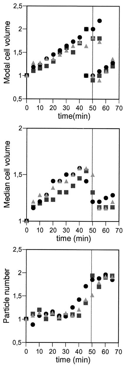
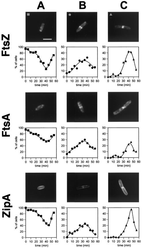
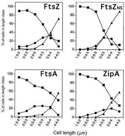
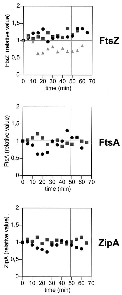
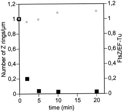
The concentration of the cell division proteins FtsZ, FtsA, and ZipA and their assembly into a division ring during the Escherichia coli B/r K cell cycle have been measured in synchronous cultures obtained by the membrane elution technique. Immunostaining of the three proteins revealed no organized structure in newly born cells. In a culture with a doubling time of 49 min, assembly of the Z ring started around minute 25 and was detected first as a two-dot structure that became a sharp band before cell constriction. FtsA and ZipA localized into a division ring following the same pattern and time course as FtsZ. The concentration (amount relative to total mass) of the three proteins remained constant during one complete cell cycle, showing that assembly of a division ring is not driven by changes in the concentration of these proteins. Maintenance of the Z ring during the process of septation is a dynamic energy-dependent event, as evidenced by its disappearance in cells treated with sodium azide.


Figures

FIG. 1.
FIG. 1.
Degree of synchrony of E. coli B/r K cultures obtained by the membrane elution method. The quality of the synchronization was monitored by measuring cell numbers and cell volumes with a Coulter counter and a Coulter channel analyzer. The modal and median cell volume and the number of cells are shown relative to the initial values. Typical cell numbers for the first point were 1 · 106 to 2 · 106 cells/ml in a volume of 10 ml. The gray vertical lines mark the doubling time for nonsynchronous cultures of this strain under the same experimental conditions. Each symbol represents results for an individual experiment.
FIG.2.
FIG.2.
Immunofluorescence microscopy showing the three different cell types after staining with the corresponding antisera and the fraction of cells showing each of the three patterns of localization of FtsZ, FtsA, and ZipA during one complete cell cycle. Cells taken at different times from the synchronous cultures were fixed and analyzed by immunofluorescence microscopy using specific antibodies. One hundred cells were analyzed for each time point. (A) Newborn cells showed no rings. (B) The rings were seen first as two fluorescent dots at both sides of the cell, i.e., the open ring. (C) Shortly before division, the ring is seen as a sharp and continuous image, i.e., the closed ring. Note the different scales in the y axes. The bar in panel A represents 2 μm and applies to all panels.
FIG. 3.
FIG. 3.
Cell types as a function of cell length. Cell length was measured for all the cells of the experiments shown in Fig. 2 and for a steady-state exponentially growing, nonsynchronous culture (FtsZNS). Then the frequencies of the three cell types identified by immunofluorescence were plotted as a function of cell length. There was a small fraction of cells (less than 2% of the population) longer than 5 μm; these were mostly cells without rings that were considered anomalous and were not included. Squares, cells without rings; circles, cells with open rings; triangles, cells with closed rings.
FIG. 4.
FIG. 4.
Relative levels of FtsZ, FtsA, and ZipA during one complete cell cycle. Samples from the synchronous cultures were taken at different times and analyzed by Western blotting with specific antibodies. Anti-EF-Tu was included in all the blots. The levels of FtsZ, FtsA, and ZipA were then measured relative to the EF-Tu band, and the ratios were made relative to the ratio at time zero; therefore, the levels of the three cell division proteins are expressed as relative concentrations. The symbols reflect results of three independent experiments for FtsZ (top) and results of two independent experiments for FtsA (middle) and ZipA (bottom). The gray vertical lines mark the doubling times of this strain in asynchronous cultures under these experimental conditions.
FIG. 5.
FIG. 5.
Energy dependence of FtsZ ring assembly. A steady-state exponential culture of E. coli MC1061 was grown in LB at 37°C to an optical density at 600 nm of 0.2. At time zero, sodium azide was added to a final concentration of 15 mM, and samples were taken for further analysis at the times indicated. Black squares, number of FtsZ rings per unit length relative to the initial value; gray circles, FtsZ/EF-Tu ratio relative to the initial value.

Similar articles

Cited by

References

    1. Addinall, S. G., and J. Lutkenhaus. 1996. FtsA is localized to the septum in an FtsZ-dependent manner. J. Bacteriol. 178:7167-7172. - PMC - PubMed
    1. Aldea, M., T. Garrido, J. Pla, and M. Vicente. 1990. Division genes in Escherichia coli are expressed coordinately to cell septum requirements by gearbox promoters. EMBO J. 9:3787-3794. - PMC - PubMed
    1. Bechtloff, D., B. Grünenfelder, T. Åkerlund, and K. Nordström. 1999. Analysis of protein synthesis rates after initiation of chromosome replication in Escherichia coli. J. Bacteriol. 181:6292-6299. - PMC - PubMed
    1. Bouché, F., and J. P. Bouché. 1989. Genetic evidence that DicF, a second division inhibitor encoded by the Escherichia coli dicB operon, is probably RNA. Mol. Microbiol. 3:991-994. - PubMed
    1. Dai, K., and J. Lutkenhaus. 1992. The proper ratio of FtsZ to FtsA is required for cell division to occur in Escherichia coli. J. Bacteriol. 174:6145-6151. - PMC - PubMed

Publication types

MeSH terms

LinkOut - more resources