. 2003 Jun;185(11):3400-9.
doi: 10.1128/JB.185.11.3400-3409.2003.

Coaggregation-mediated interactions of streptococci and actinomyces detected in initial human dental plaque

Affiliations

Coaggregation-mediated interactions of streptococci and actinomyces detected in initial human dental plaque

Robert J Palmer Jr et al. J Bacteriol. 2003 Jun.

Abstract

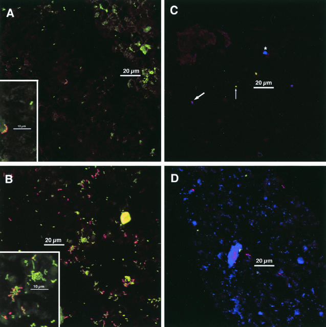
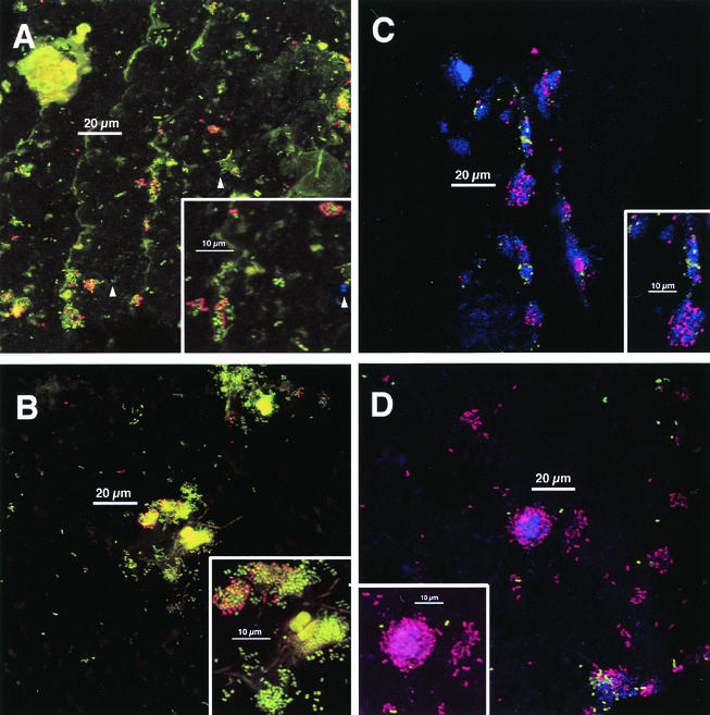
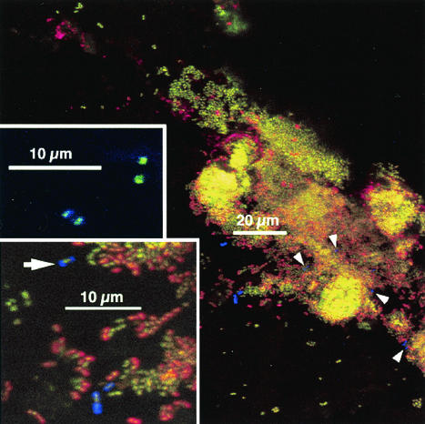
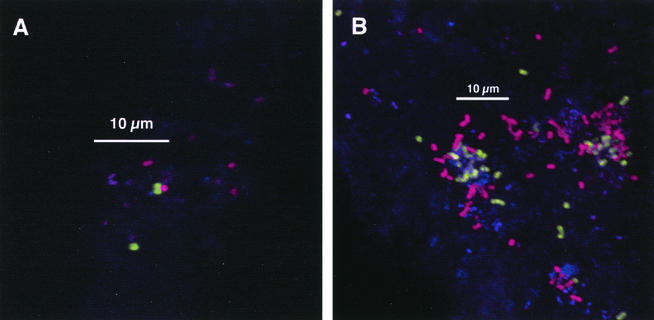
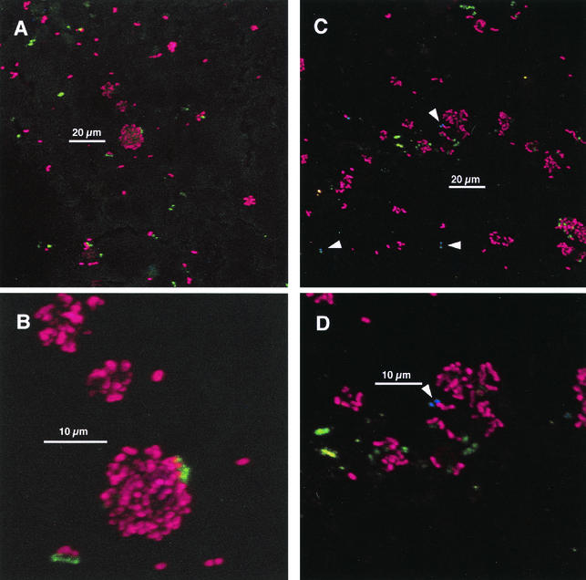
Streptococci and actinomyces that initiate colonization of the tooth surface frequently coaggregate with each other as well as with other oral bacteria. These observations have led to the hypothesis that interbacterial adhesion influences spatiotemporal development of plaque. To assess the role of such interactions in oral biofilm formation in vivo, antibodies directed against bacterial surface components that mediate coaggregation interactions were used as direct immunofluorescent probes in conjunction with laser confocal microscopy to determine the distribution and spatial arrangement of bacteria within intact human plaque formed on retrievable enamel chips. In intrageneric coaggregation, streptococci such as Streptococcus gordonii DL1 recognize receptor polysaccharides (RPS) borne on other streptococci such as Streptococcus oralis 34. To define potentially interactive subsets of streptococci in the developing plaque, an antibody against RPS (anti-RPS) was used together with an antibody against S. gordonii DL1 (anti-DL1). These antibodies reacted primarily with single cells in 4-h-old plaque and with mixed-species microcolonies in 8-h-old plaque. Anti-RPS-reactive bacteria frequently formed microcolonies with anti-DL1-reactive bacteria and with other bacteria distinguished by general nucleic acid stains. In intergeneric coaggregation between streptococci and actinomyces, type 2 fimbriae of actinomyces recognize RPS on the streptococci. Cells reactive with antibody against type 2 fimbriae of Actinomyces naeslundii T14V (anti-type-2) were much less frequent than either subset of streptococci. However, bacteria reactive with anti-type-2 were seen in intimate association with anti-RPS-reactive cells. These results are the first direct demonstration of coaggregation-mediated interactions during initial plaque accumulation in vivo. Further, these results demonstrate the spatiotemporal development and prevalence of mixed-species communities in early dental plaque.


Figures

FIG. 1.
FIG. 1.
Diagram of staining chamber. (A) Top view. The six wells are indicated by large circles with the leftmost well containing an enamel chip (square). Conduits drilled through the plastic into each well are indicated by dotted lines. Barbed connectors (black arrowheads) are threaded into each end of the conduits, and tubing is attached; the leftmost conduit shows tubing on each connector, one of which is clamped (black bar). Six screw holes (small circles) provide connections with top plate (not shown). (B) Side view. Three (of six) screws are shown passing through the transparent plastic upper plate. Six O rings on the top plate (black bars) form seals around the periphery of each well; six conduits in the lower plate are indicated by small circles.
FIG. 2.
FIG. 2.
Typical colonization after 4 h of appliance wear. Insets: electronic zoom of center region. (A and B) Staining with acridine orange (green), anti-RPS (red), and anti-type-1 (blue). (A) Sparse colonization. Chain of anti-RPS reactive cells in center; cluster of acridine orange-stained cells at lower right of scale bar. Most cells are not antibody reactive. No anti-type-1-reactive cells are visible. Marked enamel autofluorescence in upper right. Inset shows homogeneity of anti-RPS reactivity within the chain of cells. Anti-RPS reactive cells are characterized by a yellow center (green plus red) and orange-to-red edges. A pair of antibody-unreactive cells (only acridine orange staining) is visible. Other dim greenish material is debris or enamel autofluorescence. (B) Heavier colonization. Most cells are in clusters. Many anti-RPS-reactive cells. Inset shows anti-RPS-reactive cells together with acridine orange-stained cells within a mixed microcolony. (C and D) Staining with anti-DL1 (green), anti-RPS (red), and Syto 59 (blue). These images show different fields of view from a single chip. (C) Very sparse colonization. Antibody-RPS-reactive cells are purple (red plus blue; thick arrow), whereas anti-DL1-reactive cells are green (limited uptake of Syto 59; thin arrow; see text for details). A cluster of three antibody-unreactive cells (Syto 59-stained; blue; asterisk) is between scale bar and upper edge. (D) Heavier colonization. Several anti-RPS reactive cells (purple) and a few anti-DL1 reactive cells (green) are visible, but most cells are not antibody reactive (blue). Debris or enamel fluorescence (large blue regions) is visible. Maximum projection images of simultaneous-acquisition three-channel confocal stacks. Low-magnification images are shown; dimensions are 158 μm on a side and 25,000-μm2 total area (approximately 1/10 of the total chip area).
FIG. 3.
FIG. 3.
Typical colonization after 8 h of appliance wear. Staining as done for Fig. 2. Panels A, B, and D are 8-h plaque samples that correspond directly (i.e., are from the same volunteer in the same experiment) to the 4-h samples shown in Fig. 2. Insets: electronic zoom of center region. (A) Colonial nature of plaque accumulation is apparent. Two anti-type-1-reactive cells are visible (arrowheads). Inset shows heterogeneity of anti-RPS reactivity within colonies and a single anti-type-1 reactive cell (arrowhead). (B) Long, weakly fluorescent rods and large, highly fluorescent cocci (center) are visible. No anti-type-1-reactive cells are visible. Inset confirms heterogeneity of anti-RPS reactivity within colonies. (C) Colonial nature of accumulation and heterogeneity of anti-RPS-reactive cells (purple) as well as of anti-DL1-reactive cells (green) within single colonies is apparent. Inset confirms heterogeneity of antibody reactivity within colonies; three groups of cells occur (anti-DL1 reactive, anti-RPS reactive, and antibody unreactive). (D) Colony composed of anti-RPS-reactive cells, anti-DL1-reactive cells, and antibody-unreactive cells is seen in lower right. Inset shows mixed colony of anti-RPS-reactive cells and Syto 59-stained cells; anti-DL1-reactive cells (green) are in close proximity to anti-RPS-reactive cells as well as in more solitary positions.
FIG. 4.
FIG. 4.
Antibodies directed against actinomyces fimbriae are heterogeneously distributed on the cell surface, and anti-fimbria-reactive cells are infrequent on chips. Main image and large inset (zoom of central region of main image) show 8-h-old plaque stained with acridine orange (green), anti-RPS (red), and anti-type-1 (blue). Arrow in large inset indicates a cell for which anti-type-1 reactivity (blue) is concentrated at the poles; note nucleic acid stain (green) between the two blue spots. Three other anti-type-1 reactive cells (below scale bar near lower edge) display less heterogeneity. All four of these cells are visible beneath the scale bar in the main image. Arrowheads in main image point to additional anti-type-1-reactive cells that are difficult to discern because of other biomass. The location of these cells was confirmed by examining only the blue channel of the RGB image (representing output of the far-red confocal channel; Alexa 647-conjugated anti-type-1). Small inset is from a different field of view and was stained with anti-type-2 (green), anti-RPS (red), and anti-type-1 (blue). Polar localization of antifimbrial antibodies in the absence of nucleic acid stain results in two pairs of dots (contrast with cell at arrow in large inset); each pair defines a single cell. Each cell is reactive with both antibodies; the leftmost cell has more anti-type-1 reactivity (blue dominates over green) than the cell at right (green dominates over blue).
FIG. 5.
FIG. 5.
Unambiguous interactions of at least two coccoid genotypes. Staining with anti-DL1 (green), anti-RPS (red), and Syto 59 (blue). (A) Anti-DL1-reactive cells in association with an anti-RPS-reactive cell in 4-h-old plaque. (B) Interaction of anti-DL1-reactive cells, anti-RPS-reactive cells, and antibody-unreactive cells in 8-h-old plaque.
FIG. 6.
FIG. 6.
Interaction between anti-RPS-reactive cells and anti-type-2-reactive cells in 8-h-old plaque. Staining is with anti-type-2 (green), anti-RPS (red), and anti-type-1 (blue). (A and C) Low-magnification views. (B and D) Electronic zoom of central regions for panels A and C. Most actinomyces cells stained primarily with anti-type-2; arrowheads in panels C and D indicate cells for which binding of anti-type-1 predominated.

Similar articles

Cited by

References

    1. Becker, M. R., B. J. Paster, E. J. Leys, M. L. Moeschberger, S. G. Kenyon, J. L. Galvin, S. K. Boches, F. E. Dewhirst, and A. L. Griffen. 2002. Molecular analysis of bacterial species associated with childhood caries. J. Clin. Microbiol. 40:1001-1009. - PMC - PubMed
    1. Buswell, C. M., Y. M. Herlihy, P. D. Marsh, C. W. Keevil, and S. A. Leach. 1997. Coaggregation amongst aquatic biofilm bacteria. J. Appl. Microbiol. 83:477-484.
    1. Cisar, J. O., M. J. Brennan, and A. L. Sandberg. 1985. Lectin-specific interaction of Actinomyces fimbriae with oral streptococci, p. 159-163. In S. E. Mergenhagen and B. Rosan (ed.), Molecular basis of oral microbial adhesion. American Society for Microbiology, Washington, D.C.
    1. Cisar, J. O., S. H. Curl, P. E. Kolenbrander, and A. E. Vatter. 1983. Specific absence of type 2 fimbriae on a coaggregation-defective mutant of Actinomyces viscosus T14V. Infect. Immun. 40:759-765. - PMC - PubMed
    1. Cisar, J. O., V. A. David, S. H. Curl, and A. E. Vatter. 1984. Exclusive presence of lactose-sensitive fimbriae on a typical strain (WVU45) of Actinomyces naeslundii. Infect. Immun. 46:453-458. - PMC - PubMed

MeSH terms

Substances

LinkOut - more resources