

Interaction between sympathetic nerve activation and muscle fibre contraction in resistance vessels of hamster retractor muscle
- PMID: 12754308
- PMCID: PMC2343056
- DOI: 10.1113/jphysiol.2003.038984
Interaction between sympathetic nerve activation and muscle fibre contraction in resistance vessels of hamster retractor muscle
Abstract
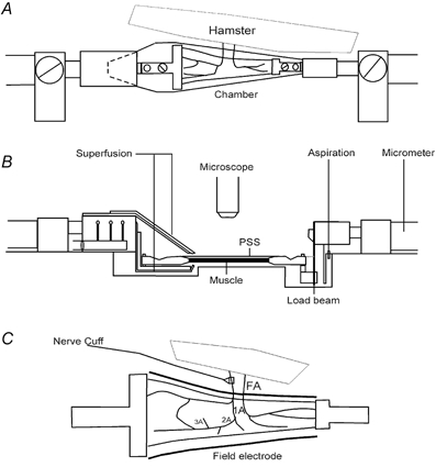
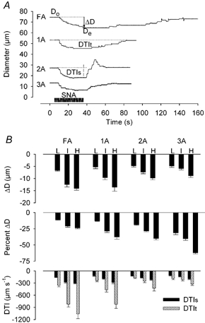
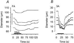
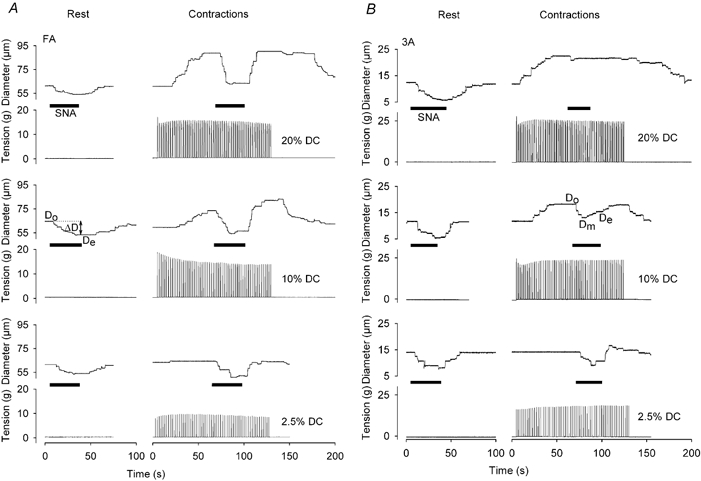
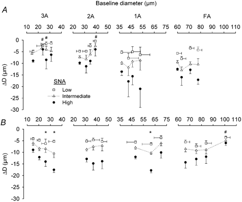
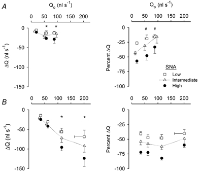
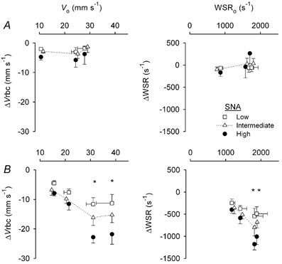
The interaction between skeletal muscle contraction and sympathetic nerve activation (SNA) on blood flow during exercise has remained ambiguous due to indirect estimates of vasomotor control. In the hamster retractor muscle (n=54), interactions between three levels of SNA (approximately 3, 6 and 12 Hz) and of contractile activity (2.5, 10 and 20 % duty cycle) were studied in feed arteries (FA) and first- (1A), second- (2A), and third-order (3A) arterioles using intravital microscopy. During functional dilatation with rhythmic muscle contractions, sympathetic vasoconstriction was sustained in FA and 1A but impaired in 2A and 3A (P<0.05), where vessels 'escaped' from responding to SNA. To account for changes in baseline diameter and blood flow during contractions, vasodilatation was induced passively (2-3 levels) in resting muscles with papaverine or sodium nitroprusside. Compared to functional dilatation, the range of passive dilatation was similar in 3A and progressively greater in 2A, 1A and FA. With passive dilatation, SNA responses were sustained in 2A and increased with baseline diameter in 3A. Blood flow through FA (rest, approximately 20 nl s(-1)) increased approximately 5-fold during contractile activity and approximately 10-fold during passive dilatation. Absolute flow reductions (nl s(-1)) with SNA increased during contractile activity and during passive dilatation; relative flow reductions were impaired during functional dilatation (P<0.05) and remained constant during passive dilatation. Thus, SNA can restrict blood flow to exercising muscle by constricting FA and 1A while dilatation prevails in 2A and 3A. Such concerted interaction will promote oxygen extraction when blood flow is restricted to maintain arterial pressure.
Figures
Comment in
-
Having it both ways? Vasoconstriction in contracting muscles.J Physiol. 2003 Jul 15;550(Pt 2):333. doi: 10.1113/jphysiol.2003.044628. Epub 2003 May 30. J Physiol. 2003. PMID: 12777450 Free PMC article. No abstract available.
Similar articles
-
Sympathetic nerves inhibit conducted vasodilatation along feed arteries during passive stretch of hamster skeletal muscle.J Physiol. 2003 Oct 1;552(Pt 1):273-82. doi: 10.1113/jphysiol.2003.046284. Epub 2003 Aug 1. J Physiol. 2003. PMID: 12897176 Free PMC article.
-
Integration of blood flow control to skeletal muscle: key role of feed arteries.Acta Physiol Scand. 2000 Apr;168(4):511-8. doi: 10.1046/j.1365-201x.2000.00703.x. Acta Physiol Scand. 2000. PMID: 10759588 Review.
-
Muscle length directs sympathetic nerve activity and vasomotor tone in resistance vessels of hamster retractor.Circ Res. 1996 Sep;79(3):551-9. doi: 10.1161/01.res.79.3.551. Circ Res. 1996. PMID: 8781488
-
Rapid dilation of arterioles with single contraction of hamster skeletal muscle.Am J Physiol Heart Circ Physiol. 2006 Jan;290(1):H119-27. doi: 10.1152/ajpheart.00197.2005. Epub 2005 Aug 12. Am J Physiol Heart Circ Physiol. 2006. PMID: 16100250
-
Neural control of muscle blood flow during exercise.J Appl Physiol (1985). 2004 Aug;97(2):731-8. doi: 10.1152/japplphysiol.00076.2004. J Appl Physiol (1985). 2004. PMID: 15247201 Review.
Cited by
-
Exercise hyperaemia: is anything obligatory but the hyperaemia?J Physiol. 2007 Sep 15;583(Pt 3):855-60. doi: 10.1113/jphysiol.2007.135889. Epub 2007 Jul 19. J Physiol. 2007. PMID: 17640934 Free PMC article. Review.
-
Regional heterogeneity of α-adrenoreceptor subtypes in arteriolar networks of mouse skeletal muscle.J Physiol. 2010 Nov 1;588(Pt 21):4261-74. doi: 10.1113/jphysiol.2010.194993. J Physiol. 2010. PMID: 20807785 Free PMC article.
-
Theoretical model of metabolic blood flow regulation: roles of ATP release by red blood cells and conducted responses.Am J Physiol Heart Circ Physiol. 2008 Oct;295(4):H1562-71. doi: 10.1152/ajpheart.00261.2008. Epub 2008 Aug 8. Am J Physiol Heart Circ Physiol. 2008. PMID: 18689501 Free PMC article.
-
ASIC3 contributes to the blunted muscle metaboreflex in heart failure.Med Sci Sports Exerc. 2015 Feb;47(2):257-63. doi: 10.1249/MSS.0000000000000415. Med Sci Sports Exerc. 2015. PMID: 24983337 Free PMC article.
-
Aging alters reactivity of microvascular resistance networks in mouse gluteus maximus muscle.Am J Physiol Heart Circ Physiol. 2014 Sep 15;307(6):H830-9. doi: 10.1152/ajpheart.00368.2014. Epub 2014 Jul 11. Am J Physiol Heart Circ Physiol. 2014. PMID: 25015968 Free PMC article.
References
-
- Anderson KM, Faber JE. Differential sensitivity of arteriolar alpha 1- and alpha 2-adrenoceptor constriction to metabolic inhibition during rat skeletal muscle contraction. Circ Res. 1991;69:174–184. - PubMed
-
- Boegehold MA, Johnson PC. Periarteriolar and tissue PO2 during sympathetic escape in skeletal muscle. Am J Physiol. 1988a;254:H929–936. - PubMed
-
- Boegehold MA, Johnson PC. Response of arteriolar network of skeletal muscle to sympathetic nerve stimulation. Am J Physiol. 1988b;254:H919–928. - PubMed
Publication types
MeSH terms
Grants and funding
LinkOut - more resources
Full Text Sources
Research Materials