. 2003 May 27;42(20):6090-8.
doi: 10.1021/bi0342416.

Purification and characterization of a transmembrane domain-deleted form of lecithin retinol acyltransferase

Affiliations

Purification and characterization of a transmembrane domain-deleted form of lecithin retinol acyltransferase

Dean Bok et al. Biochemistry. .

Abstract

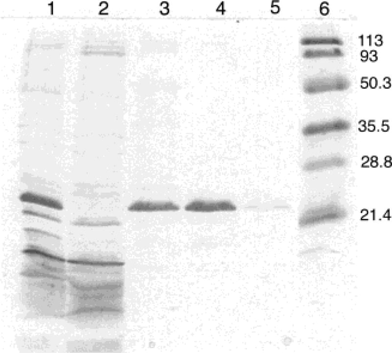
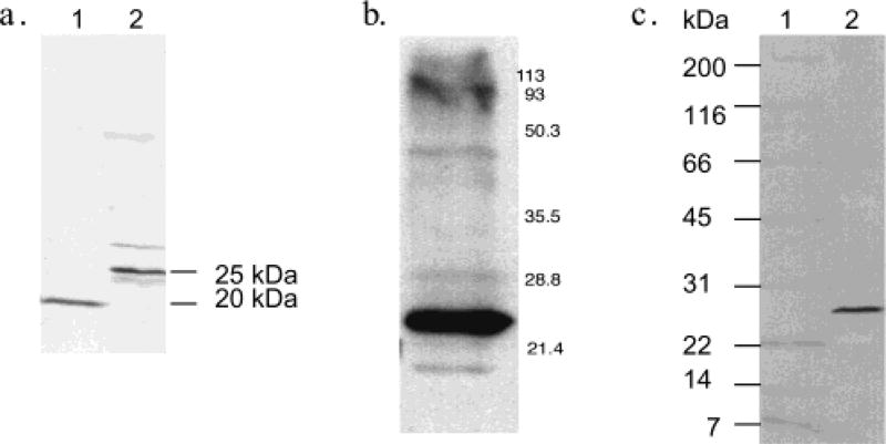
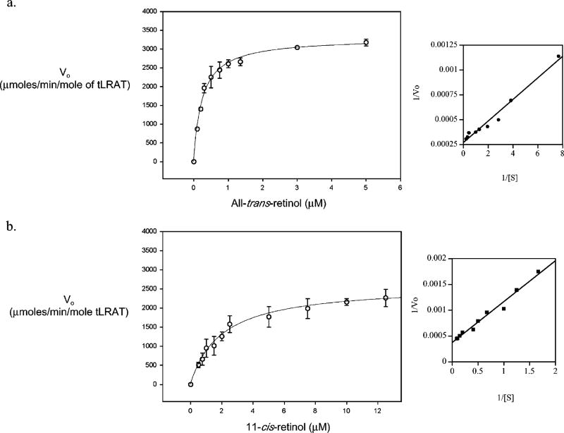
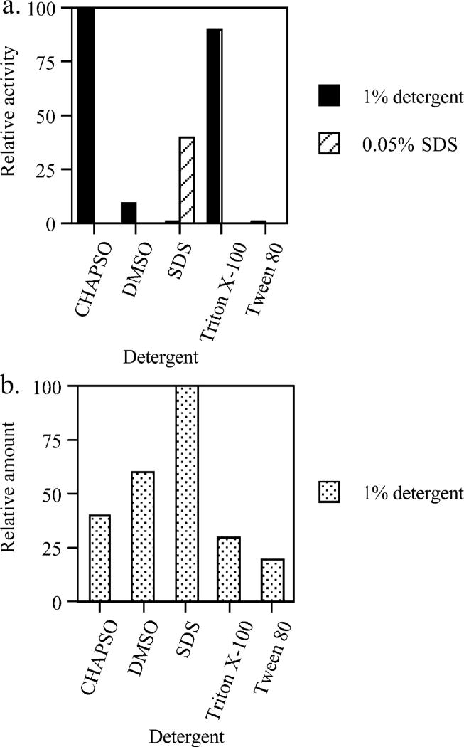
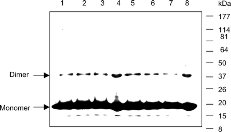
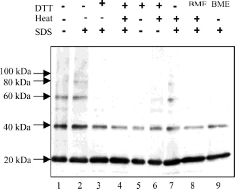
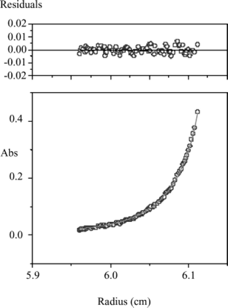
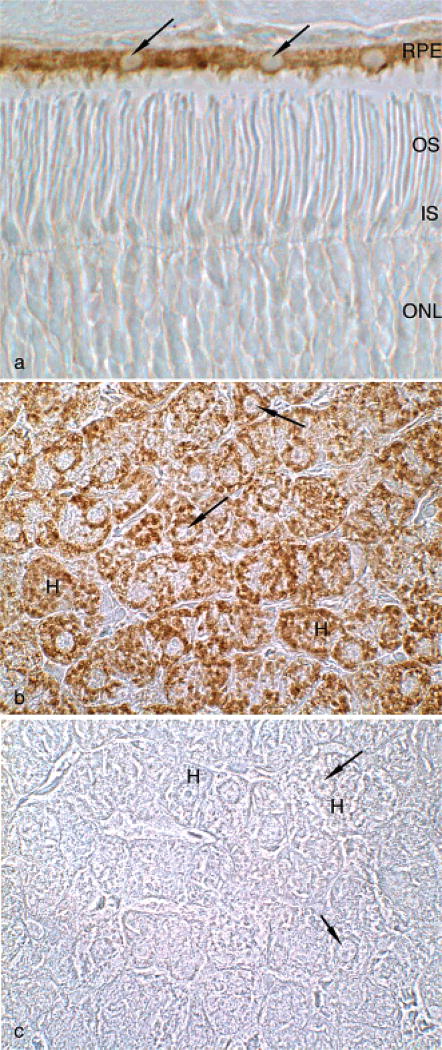
Lecithin retinol acyltransferase (LRAT) catalyzes the esterification of all-trans-retinol into all-trans-retinyl ester, an essential reaction in the vertebrate visual cycle. Since all-trans-retinyl esters are the substrates for the isomerization reaction that generates 11-cis-retinoids, this esterification reaction is essential in the operation of the visual cycle. In addition, LRAT is the founder member of a series of proteins, which are of novel sequence and have unknown functions. Native LRAT is an integral membrane protein and has never been purified. To obtain a pure LRAT, the N- and C-transmembrane termini were deleted and replaced with a poly His tag for the purpose of purification. This truncated form of LRAT, referred to as tLRAT, has been expressed in bacteria and fully purified. tLRAT is catalytically active and processes all-trans-retinol at least 10-fold more efficiently than 11-cis-retinol, the precursor to the visual chromophore. While tLRAT can be robustly expressed in bacteria, it requires detergent for extraction, as the enzyme still contains hydrophobic domains, which may interact. Indeed, tLRAT can oligomerize and forms dimers. Native LRAT also forms functional homodimers. These studies pave the way for the preparation of large-scale amounts of pure tLRAT for further mechanistic and structural studies.


Figures

Figure 1
Figure 1
Silver stained SDS–PAGE of tLRAT purified on Ni column in SDS. Lane 1, cell extract; lane 2, Ni column flowthrough; lane 3, pH 6.0 elution; lane 4, pH 5.3 elution; lane 5, pH 4.0 elution; and lane 6, molecular weight markers.
Figure 2
Figure 2
(a) Western blot of the purified tLRAT (lane 1) and LRAT (lane 2) from the crude RPE. The membrane was probed with anti-LRAT peptide polyclonal antibody (1:4000), anti-rabbit Ig linked horseradish peroxidase (1:8000), and ECL reagent. (b) Western blot of tLRAT of a bacterial lysate reacted with anti-LRAT protein antibody. (c) Western blot of LRAT in the RPE (lane 2). Molecular weight markers (lane 1) are 200, 116, 66, 45, 31, 22, 14, and 7 kDa, respectively.
Figure 3
Figure 3
Steady-state kinetics of tLRAT using all-trans-retinol and 11-cis-retinol as substrates. Esterification activity of tLRAT was monitored by following the formation of retinyl esters using tritium labeled all-trans-retinol and 11-cis-retinol along with DPPC. 5–10 µg of purified tLRAT was used in 100 mM Tris-HCl (100 µL) at pH 8.3 containing 220 µM DPPC, 0.6% BSA, 1 mM EDTA, 2 mM DTT, 0.1% CHAPSO, and 0.2 µM of the retinol. (a) The initial velocity of all-trans-retinyl ester formation reaction and subsequent Lineweaver–Burk plot. (b) The initial velocity of 11-cis-retinyl ester formation reaction and subsequent Lineweaver–Burk plot.
Figure 4
Figure 4
Relative activity and solibilization of tLRAT in the presence of different agents. (a) tLRAT was extracted with each detergent for 2–4 h at 4 °C, and all-trans-retinol esterification activity was measured as described in the Experimental Procedures. Relative amounts of tLRAT extracted were determined by SDS–PAGE/Western blot. In the absence of added detergent, minimum tLRAT activity (20%) was observed in the 13 800g centrifuged supernatant, while no activity (<1%) was observed in the 265 000g sample. (b) The relative extraction amounts and the catalytic activities showed about 10% variation depending on the mechanical force used (e.g., magnetic stirring vs gentle shaking). The values are the averages of at least five determinations.
Figure 5
Figure 5
tLRAT homodimer analysis by mass spectrometry. Proteins were visualized by Coomassie blue staining after SDS–PAGE. Hisx6 tagged truncated LRAT (tLRAT) purified by Ni2+ column using an imidazole gradient elution buffer (10–500 mM). SDS–PAGE gel, in each lane, 40 µL (80 µg) of sample was loaded. Lanes 1–3 and 5–7, tLRAT in SDS (2%) + DTT (5 mM) + heating; lanes 4 and 8, tLRAT in SDS (2%) without DTT/heating. The 20 and 40 kDa bands were excised, trypsin digested, and analyzed by electrospray tandem mass spectrometry showing 13 (20 kDa) and 11 (40 kDa) LRAT peptides, respectively.
Figure 6
Figure 6
Purified tLRAT oligomer formation. Purified tLRAT was incubated 45 min in the reducing condition (DTT) and/or detergent (SDS). Protein was visualized by Western blot analysis. Lane 1, native sample buffer only; lane 2, SDS (2%); lane 3, SDS (2%) + DTT(10 mM); lane 4, SDS (2%) + DTT (5 mM) + heating; lane 5, DTT (5 mM); lane 6, DTT (5 mM) + heating (2 min); lane 7, SDS (2%) + heating (2 min); lane 8, SDS (2%) + BME (10%) + heating (2 min); and lane 9, SDS (2%) + BME (10%).
Figure 7
Figure 7
Sedimentation equilibrium analysis of tLRAT. tLRAT interaction analysis was performed in a Beckman Optima XL-A analytical ultracentrifuge using a 4-hole rotor and six channel cell. A solution of 100 µL of tLRAT (15 µM) in a buffer containing 50 mM sodium phosphate, pH 7.4, 100 mM NaCl, and 10 mM DTT is shown as absorbance vs radial position at equilibrium at 20 °C and 45 000 rpm. The scans were taken at 280 nm and 0.01 mm radial step resolution. Analysis of the scanned data was performed with the Origin 3.78 program. The raw data (circles) and the global fit of a monomer–dimer equilibrium model (lines) are shown.
Figure 8
Figure 8
Immunocytochemistry of bovine retina and liver using affinity purified anti tLRAT antibodies. (a) In the retina, staining is limited to the retinal pigment epithelium (RPE). Retinal photoreceptor outer segments (OS), inner segments (IS), and outer nuclear layer (ONL) are shown for comparison. (b) Staining in the liver is observed in hepatocytes (H). The nuclei of the RPE and hepatocytes (arrows) are negative, consistent with the putative location of LRAT in the smooth endoplasmic reticulum of cells. (c) Control section of liver in which immune IgG was replaced with preimmune IgG. × 270.

Similar articles

Cited by

References

    1. MacDonald PN, Ong DE. J. Biol. Chem. 1988;263:12478–12482. - PubMed
    1. Barry R, Cañada FJ, Rando RR. J. Biol. Chem. 1989;264:9231–9238. - PubMed
    1. Saari JC, Bredberg DL. J. Biol. Chem. 1989;264:8636–8640. - PubMed
    1. Ruiz A, Winston A, Lim Y-H, Gilbert BZ, Rando RR, Bok D. J. Biol. Chem. 1999;274:3834–3841. - PubMed
    1. MacDonald PN, Ong DE. Biochem. Biophys. Res. Commun. 1988;156:157–163. - PubMed

Publication types

MeSH terms