. 2003 May 26;161(4):757-68.
doi: 10.1083/jcb.200302151. Epub 2003 May 19.

The Caenorhabditis elegans vab-10 spectraplakin isoforms protect the epidermis against internal and external forces

Affiliations

The Caenorhabditis elegans vab-10 spectraplakin isoforms protect the epidermis against internal and external forces

Julia M Bosher et al. J Cell Biol. .

Abstract

Morphogenesis of the Caenorhabditis elegans embryo is driven by actin microfilaments in the epidermis and by sarcomeres in body wall muscles. Both tissues are mechanically coupled, most likely through specialized attachment structures called fibrous organelles (FOs) that connect muscles to the cuticle across the epidermis. Here, we report the identification of new mutations in a gene known as vab-10, which lead to severe morphogenesis defects, and show that vab-10 corresponds to the C. elegans spectraplakin locus. Our analysis of vab-10 reveals novel insights into the role of this plakin subfamily. vab-10 generates isoforms related either to plectin (termed VAB-10A) or to microtubule actin cross-linking factor plakins (termed VAB-10B). Using specific antibodies and mutations, we show that VAB-10A and VAB-10B have distinct distributions and functions in the epidermis. Loss of VAB-10A impairs the integrity of FOs, leading to epidermal detachment from the cuticle and muscles, hence demonstrating that FOs are functionally and molecularly related to hemidesmosomes. We suggest that this isoform protects against forces external to the epidermis. In contrast, lack of VAB-10B leads to increased epidermal thickness during embryonic morphogenesis when epidermal cells change shape. We suggest that this isoform protects cells against tension that builds up within the epidermis.


Figures

Figure 1.
Figure 1.
vab-10 mutants display elongation and body morphology defects. Differential interference contrast micrographs of terminal-stage vab-10 mutants. (A) Wild-type twofold embryo (mid-embryogenesis). (B) vab-10(h1356) embryo; the body (demarcated by arrowheads) failed to elongate. (C) vab-10A(ju281) embryo with a localized detachment of the epidermis from the cuticle (arrow); all vab-10A(ju281) embryos raised at 20°C and 79% of those raised at 25°C (n = 149) elongated 2.5-fold like this embryo, and occasionally hatched to generate kinked and paralyzed larvae, whereas 21% of those raised at 25°C looked like h1356 embryos. (D) vab-10A(RNAi) embryo; 95% of these embryos (n = 135) resembled h1356 embryos. (E) Arrested L1 vab-10B(mc44) larva (65%, n = 403, could hatch), and (F) hatching L1 vab-10B(RNAi) larva; the body morphology is very irregular. Embryos laid after eliciting an RNAi response against vab-10A– or vab-10B–specific exons are denoted vab-10A(RNAi) or vab-10B(RNAi). Here (as in Figs. 4, 6, and 8), dorsal is up, anterior is left, and bars represent 10 μm.
Figure 2.
Figure 2.
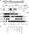
vab-10 encodes two distinct plakins. (A) The first two lines represent the chromosomal region where vab-10 maps and shows the approximate (dotted areas) endpoints of the deficiencies hDf17 and hDf16 used to refine vab-10 position. vab-10 spreads over two overlapping cosmids (F53B8 and ZK1151) and a yeast artificial chromosome (Y47H9B). The first 15 exons (dark gray boxes, corresponding to a predicted gene known as ZK1151.1) are common to vab-10A and vab-10B isoforms; exons 16–17 (medium gray boxes, corresponding to a predicted gene known as ZK1151.3) are unique to vab-10A isoforms, exons 18–32 (light gray boxes, corresponding to a predicted gene known as ZK1151.2) are unique to vab-10B isoforms. Numbered exons (arrowheads) can be alternatively spliced (see Fig. S1). Arrows mark the positions of vab-10 mutations as follows: h1356 is a G to A transition in the GT consensus donor splice site of intron 10, leading to a premature stop codon downstream; ju281 is a G to A transition (nucleotide 20242 of ZK1151) changing the Gly1560 of the longest VAB-10A isoform into a Glu; e698 is a G to A transition (nucleotide 19925 of ZK1151) changing the Pro1666 of the longest VAB-10A isoform into a Ser; mc44 is a 1033-nucleotide deletion (spanning nucleotides 2636–1605 of ZK1151) truncating VAB-10B isoforms after residue 3515. Shown below are the functional domains predicted by the SMART program (http://smart.embl-heidelberg.de) in VAB-10A, VAB-10B, and the two most closely related vertebrate plakins. ABD, actin-binding domain (exons encoding the ABD are surrounded by a dotted box); CH, calponin homology; SH3, Src-homology domain 3; SR, spectrin repeat; PR, plectin repeat; GAR, growth-arrest protein 2-related homology; EF-hand, calcium-binding motif. (B) Complementation tests among vab-10 alleles bearing on at least 100 individuals of each genotype. Only the most common phenotypes are mentioned (<C, 1.7F, 2F, 2.5F: arrest at the comma, 1.7-fold, twofold, and 2.5-fold stages, respectively; L1: L1 arrest; Vab, variably abnormal; WT, wild-type; ND, not determined).
Figure 3.
Figure 3.
Characterization of four VAB-10–specific antibodies. (Top) Positions and names (Prot) of the peptides (horizontal double arrows; domain symbols are as in Fig. 2 A) used to raise antibodies (Ab), and of the epitope recognized by the mAb MH5 (vertical arrow). (Bottom) Western blots of total worm extracts probed with immunopurified antibodies (Ab) in the absence (−) or in the presence of a competing protein (C.P.). Competition with 4fr, kh2, and kh3 recombinant peptides were effective only with the cognate antibodies (e.g., 4fr did not compete interaction with mAb MH5).
Figure 4.
Figure 4.
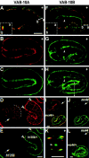
VAB-10 antibodies recognize regions of epidermis–muscle contact. Confocal projections of embryos stained with antibodies against VAB-10A (4F2: A, C, and E; MH5: B and D) or VAB-10B (K22: F, G, I, and J; K32: H, K, and L), and with the mAbs MH46 (recognizes myotactin at the basal epidermal membrane in regions of muscle contact; A, F, and I), NE8/4C6 (K, muscle-specific), or MH27 (D, adherens junction-specific; staining with mAbs is in red). (A and F) Early wild-type comma embryos; VAB-10A and VAB-10B staining is detected at the basal (b) and apical (a) membranes as further shown in optical cross sections along the apico–basal axis at the level of arrows (3× insets surrounded by a dotted line). (B and C) Wild-type embryo revealing three muscle–epidermis contact areas. (D and E) vab-10(h1356) embryo (arrow) and a neighboring heterozygous sibling (right); the VAB-10A signal is absent in the h1356 mutant embryo even at a stage when muscles are not yet functional (MH27 staining reveals that fusion between dorsal epidermal cells (dots), which normally precedes the first muscle contraction, did not occur yet; arrowheads show two areas of muscle contact in the control). (G and H) Wild-type embryos revealing two muscle–epidermis contact areas, the pharynx and the intestine (arrowhead). (I and J) vab-10B(mc44) embryo (arrow) and a neighboring heterozygous sibling (bottom); there is a very faint residual staining with K22 antibodies. (K) Z-optical projection through the entire stack of images for the embryo in H at the level of the white bar; VAB-10B is found above muscles (in red) and in the intestine (arrowhead). (L) vab-10B(mc44) embryo (arrow) and a neighboring heterozygous sibling (bottom); there is essentially no staining with K32 antibodies. Bars, 10 μm.
Figure 5.
Figure 5.
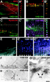
VAB-10A and VAB-10B form alternating circumferential bands in the larval epidermis. (Top) Confocal projections (A, B, C, D, and E–E′′), and optical section through the apico–basal axis along the area marked with a double arrow (A′, B′, C′, and D′) after staining wild-type adult animals (A/A′, D/D′, and E′/E′′) or L1 larvae (B/B′ and C/C′); mAb staining is in red. (A/A′) VAB-10A pAbs and myosin heavy chain–specific mAb 5.6.1.1; note the respective orientations (A) and thickness (A′) of sarcomeres and FOs. (B/B′) VAB-10A pAbs (green) and mAb MH4 (IFs; red); both proteins colocalize. (C/C′) VAB-10A pAbs and mAb MH46 (myotactin); these proteins do not generally colocalize. (D/D′) mAb MH5 (VAB-10A; green) and VAB-10B K22 pAbs (violet); these proteins do not colocalize and VAB-10B extends slightly further than VAB-10A. (E–E′′) Differential interference contrast picture (E) of the animal immunostained with VAB-10B K22 pAbs (E′); the merged image (E′′) shows that VAB-10B is found at the furrows separating annuli (arrowheads; due to the permeabilization treatments, their morphology is rather poor). The K32 antiserum revealed the same pattern, but it was much fainter (not depicted). Bar, 10 μm (A and D), 2.5 μm (B and C), or 5 μm (E). (Bottom) Immunogold- labeled micrographs showing the positions of VAB-10A (F and G), and VAB-10B (H) obtained with 4F2 and K22 antibodies, respectively. In the epidermis, VAB-10A (but not VAB-10B) is enriched in FOs (arrowheads, gold particles; white arrows, dense bodies; F and G are from different sections). The specificity of staining is indicated by the signal-to-noise ratio measured by counting gold beads in sections stained with 4F2 or K22 primary antibodies, versus in control sections without primary antibody. 4F2: epidermis, 36.7 ± 4.6, and muscle, 0.9 ± 0.2; K22: epidermis, 4.3 ± 0.5, and muscle, 3.7 ± 0.6 (n = 2). Bars: 500 nm (F and H) and 100 nm (G).
Figure 6.
Figure 6.
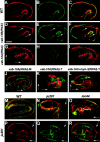
Integrity and attachment defects in vab-10 mutants. Confocal projections after staining mid-stage (A–J and P–R) or terminal-stage embryos (K–O) with the muscle-specific mAb NE8/4C6 (red) and VAB-10A 4F2 antibodies (green; A–I and M–O) or VAB-10B K22 antibodies (green; J–L and P–R); C, F, I, and R correspond to merged images of A and B, D and E, G and H, and P and Q, respectively. (A–C) Wild-type embryo; muscles are adjacent to the epidermis and the outer surface. (D–F) vab-10B(RNAi) embryo; the epidermal layer is enlarged, although muscles remain adjacent to the basal layer of the epidermis (arrows). (G–K) vab-10A(RNAi) embryos; muscles always occupied normal positions at mid-embryogenesis extending to the tip of the head and tail (G and J, arrows), but subsequently detached and collapsed to the center of the embryo in 90% of the cases (K, n = 70). (L) Simultaneous RNAi against vab-10A and myo-3 (myosin heavy chain gene); muscles remained attached in the head and tail in 74% of the embryos (n = 69). (M) Wild-type embryo. (N) vab-10A(ju281) and (O) vab-10B(mc44) embryos; VAB-10A is almost absent where muscles have detached (arrows), which occurred in >65% vab-10B(mc44) embryos and 81% vab-10A(ju281) embryos (n > 50). (P–R) vab-10A(ju281) embryo; in some areas (arrows) muscles have detached, causing localized loss of VAB-10B staining (a control embryo costained for K32 and NE8/4C6 antibodies is shown in Fig. 4, H and K).
Figure 7.
Figure 7.
VAB-10A–deficient mutants have nonfunctional FOs, VAB-10B–deficient mutants have an enlarged epidermis. EM analysis of cuticle–epidermis–muscle attachments of wild-type (A), vab-10A(RNAi) (B and F), vab-10A(ju281) (C), vab-10B(RNAi) (D), and vab-10(h1356) (E) embryos. B corresponds to a twofold enlargement of the area boxed in F. Sections are longitudinal to visualize FOs. The wild-type embryo (A) was at a stage when the muscle mass is not yet strongly developed (the fixation procedure used does not allow to visualize IFs). FOs were less numerous and/or not fully functional in VAB-10A–deficient embryos (B, C, and F), but appeared essentially normal in VAB-10B–deficient embryos (D), whereas the epidermal thickness was irregular and significantly increased in VAB-10B–deficient embryos (D); note that vab-10(h1356) embryos combined both defects (E). Black arrowheads, FO plaque at the apical plasma membrane; semi-open arrowheads, FO plaque at the basal plasma membrane; white open arrows, dense bodies at the muscle plasma membrane; capital S in white oval, sarcomeres; double arrows, epidermal layer. Bars, 1 μm.
Figure 8.
Figure 8.
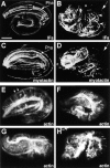
vab-10 is required to organize other FO components and actin MFs. Late-stage embryos were stained with the IF-specific mAb MH4 (A and C), the myotactin-specific mAb MH46 (C and D), or with rhodamine-conjugated phalloidin (E–H). (A and B) Wild-type pretzel embryos; the distributions of IF and myotactin are very similar to what is observed in L1 larvae (see Fig. 5, B and C; both markers are also expressed in the pharynx, see Pha). (B and D) vab-10A(RNAi) embryo; there is very little IF or myotactin left in the dorsal epidermis (arrow), and IFs tend to bundle together ventrally along what could correspond to lateral membranes (C, arrowheads). (E) Wild-type threefold stage embryo; actin MFs are evenly spaced and oriented along the circumference (arrows). (F) vab-10A(RNAi) embryo; many actin MFs are irregularly oriented. (G) vab-10B(RNAi) embryo; actin MFs were reproducibly thin and difficult to visualize. (H) vab-10(h1356) embryo; actin MFs were short or absent and randomly oriented. Bar, 10 μm.
Figure 9.
Figure 9.
Model for the distributions and roles of VAB-10 isoforms in the epidermis. Enlarged view of a muscle–epidermis–cuticle contact area in elongating wild-type (left), vab-10A (middle), and vab-10B (right) embryos. FOs correspond to the structure formed by electron-dense plaques found at the apical and basal epidermal plasma membranes (black bars) and the interconnecting IFs. Myotactin, MUP-4, and MUA-3 connect FOs to the cuticle (Cut) or the ECM separating the epidermis (Epid) from muscles (Mus). VAB-10A (pink) is an FO component (green), and VAB-10B is interspersed between FOs (pink) and coincides with the furrows separating annuli where actin MFs have also been observed (Costa et al., 1997). Myotactin bands do not fully coincide with VAB-10A bands in young larvae, but do so at later stages. VAB-10B is clearly apical and basal in young embryos, but we do not know if this is also the case later. VAB-10A absence affects FO assembly, causing the epidermis to detach from the ECM and the cuticle. VAB-10B absence causes the epidermal thickness to increase. The molecules that help maintain the distance between apical and basal plasma membranes together with VAB-10B are further discussed in the text. The respective thickness of each layer is not drawn to scale.

References

    1. Andra, K., H. Lassmann, R. Bittner, S. Shorny, R. Fassler, F. Propst, and G. Wiche. 1997. Targeted inactivation of plectin reveals essential function in maintaining the integrity of skin, muscle, and heart cytoarchitecture. Genes Dev. 11:3143–3156. - PMC - PubMed
    1. Andra, K., B. Nikolic, M. Stocher, D. Drenckhahn, and G. Wiche. 1998. Not just scaffolding: plectin regulates actin dynamics in cultured cells. Genes Dev. 12:3442–3451. - PMC - PubMed
    1. Baum, P.D., and G. Garriga. 1997. Neuronal migrations and axon fasciculation are disrupted in ina-1 integrin mutants. Neuron. 19:51–62. - PubMed
    1. Bercher, M., J. Wahl, B.E. Vogel, C. Lu, E.M. Hedgecock, D.H. Hall, and J.D. Plenefisch. 2001. mua-3, a gene required for mechanical tissue integrity in Caenorhabditis elegans, encodes a novel transmembrane protein of epithelial attachment complexes. J. Cell Biol. 154:415–426. - PMC - PubMed
    1. Brenner, S. 1974. The genetics of Caenorhabditis elegans. Genetics. 77:71–94. - PMC - PubMed

Publication types

MeSH terms

Substances