. 2003 May 26;161(4):737-47.
doi: 10.1083/jcb.200212140. Epub 2003 May 19.

Colocalization of synapsin and actin during synaptic vesicle recycling

Affiliations

Colocalization of synapsin and actin during synaptic vesicle recycling

Ona Bloom et al. J Cell Biol. .

Abstract

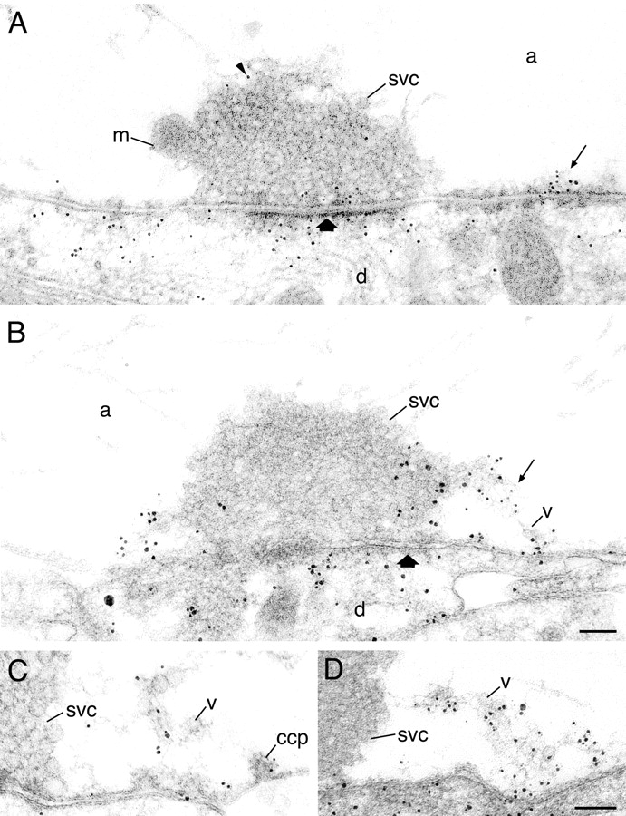
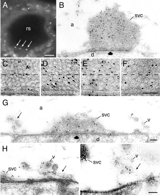
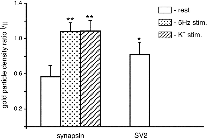
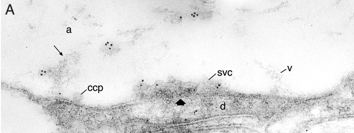
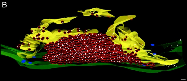
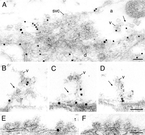
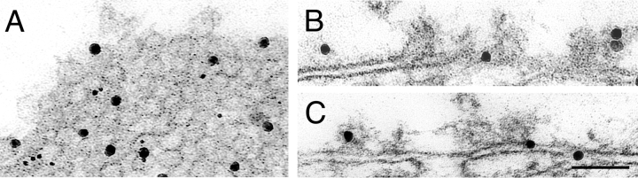
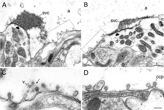
It has been hypothesized that in the mature nerve terminal, interactions between synapsin and actin regulate the clustering of synaptic vesicles and the availability of vesicles for release during synaptic activity. Here, we have used immunogold electron microscopy to examine the subcellular localization of actin and synapsin in the giant synapse in lamprey at different states of synaptic activity. In agreement with earlier observations, in synapses at rest, synapsin immunoreactivity was preferentially localized to a portion of the vesicle cluster distal to the active zone. During synaptic activity, however, synapsin was detected in the pool of vesicles proximal to the active zone. In addition, actin and synapsin were found colocalized in a dynamic filamentous cytomatrix at the sites of synaptic vesicle recycling, endocytic zones. Synapsin immunolabeling was not associated with clathrin-coated intermediates but was found on vesicles that appeared to be recycling back to the cluster. Disruption of synapsin function by microinjection of antisynapsin antibodies resulted in a prominent reduction of the cytomatrix at endocytic zones of active synapses. Our data suggest that in addition to its known function in clustering of vesicles in the reserve pool, synapsin migrates from the synaptic vesicle cluster and participates in the organization of the actin-rich cytomatrix in the endocytic zone during synaptic activity.


Figures

Figure 1.
Figure 1.
Ultrastructural localization of actin in resting and stimulated reticulospinal synapses. (A) Electron micrograph of a synapse in an unstimulated specimen labeled with an anti-actin antibody. Arrowhead indicates gold particle. Thick arrow indicates active zone. (B) Immunogold localization of actin in a synapse stimulated with action potentials at 5 Hz. Thin arrow indicates filamentous matrix in the endocytic zone. (C and D) Endocytic zones in two different stimulated synapses. Note clathrin-coated intermediates (ccp) at the presynaptic membrane in stimulated axons. m, mitochondrion; svc, synaptic vesicle cluster; v, vesicles in the filamentous matrix; d, dendrite; a, axoplasmic matrix. Bars, 100 nm.
Figure 2.
Figure 2.
Redistribution of synapsin in resting and stimulated reticulospinal synapses. (A) Transverse section of a reticulospinal axon (rs) stained with lamprey-specific synapsin antibodies using immunofluorescence. Punctate labeling (arrows) is localized close to the surface of the axon. (B) The ultrastructural localization of synapsin at rest. Gold particles are associated with the synaptic vesicle cluster. Thick arrows indicate active zone. (C–F) An increase in synapsin labeling in the area within 100 nm from the presynaptic membrane in the vesicle cluster at different states: (C) rest; (D) 5 Hz action potential stimulation; (E) high K+ stimulation; (F) control, SV2 immunolabeling at rest. (G–I) Electron micrographs of synapses stimulated by action potentials at 5 Hz for 20 min and labeled for synapsin. Thin arrow indicates filamentous matrix. Note that synapsin is associated with the filamentous cytomatrix and vesicles in the endocytic zone. svc, synaptic vesicle cluster; v, vesicles in the filamentous matrix; d, dendrite; a, axoplasmic matrix. Bars: (A) 20 μm; (F) 100 nm, also for C–E; (G) 100 nm, also for B; (I) 100 nm, also for H.
Figure 3.
Figure 3.
Quantitative evaluation of gold particle distribution at active zones at rest and during synaptic activity. The bar graph shows the ratio of gold particle densities between 0–100 nm and 100–200 nm from the active zone. At rest, n = 8; 5 Hz, n = 13, and K+ stimulation, n = 12.
Figure 4.
Figure 4.
3-D distribution of synapsin in stimulated reticulospinal synapse. (A) Electron micrograph of one serial section of a reticulospinal synapse stimulated at 5 Hz for 20 min, shown reconstructed in B. Synapsin is present on vesicles (v) in the filamentous cytomatrix (arrow). svc, synaptic vesicle cluster; v, vesicles in the filamentous matrix; d, dendrite; a, axoplasmic matrix. ccp, clathrin-coated pit. This arrow indicates active zone. (B) 3-D reconstruction of 10 serial ultrathin sections. The area occupied by actin-rich filamentous cytomatrix lateral to the vesicle cluster is shown in yellow. The plasma membrane is depicted in green. All synaptic vesicles are shown in red. Colloidal gold particles are indicated by white spheres. Clathrin-coated endocytic intermediates are shown as flat blue discs on the plasma membrane. Bar, 100 nm.
Figure 4.
Figure 4.
3-D distribution of synapsin in stimulated reticulospinal synapse. (A) Electron micrograph of one serial section of a reticulospinal synapse stimulated at 5 Hz for 20 min, shown reconstructed in B. Synapsin is present on vesicles (v) in the filamentous cytomatrix (arrow). svc, synaptic vesicle cluster; v, vesicles in the filamentous matrix; d, dendrite; a, axoplasmic matrix. ccp, clathrin-coated pit. This arrow indicates active zone. (B) 3-D reconstruction of 10 serial ultrathin sections. The area occupied by actin-rich filamentous cytomatrix lateral to the vesicle cluster is shown in yellow. The plasma membrane is depicted in green. All synaptic vesicles are shown in red. Colloidal gold particles are indicated by white spheres. Clathrin-coated endocytic intermediates are shown as flat blue discs on the plasma membrane. Bar, 100 nm.
Figure 5.
Figure 5.
Distribution of synapsin and actin in active synapses as revealed by double labeling. (A) Electron micrograph of a synapse stimulated with high K+ for 30 min. Large gold particles (enhanced 10 nm) denote actin and small (enhanced 5 nm) denote synapsin immunoreactivity. (B–D) Regions of endocytic zones, containing filamentous cytomatrix in three synapses stimulated at 5 Hz for 20 min and labeled with immunogold as in A. (E and F) Electron micrographs of endocytic regions containing clathrin-coated intermediates in synapses stimulated with high K+ for 30 min. svc, synaptic vesicle cluster; v, vesicles in the filamentous matrix; d, dendrite; a, axoplasmic matrix. Bars, 100 nm.
Figure 6.
Figure 6.
Localization of synapsin in the endocytic zone. Electron micrographs from synapses double labeled with synapsin (small particles) and SV2 (large particles) antibodies. Note an even distribution of both antigens in the vesicle cluster in A and the accumulation of only SV2 immunoreactivity on the plasma membrane and endocytic intermediates in B and C. Bar, 100 nm.
Figure 7.
Figure 7.
Disruption of the actin-rich cytomatrix by microinjection of synapsin antibodies. (A) A reticulospinal synapse in an uninjected axon subjected to high frequency stimulation at 18 Hz for 6 min. Boxed area is shown in C at higher magnification. (B) A synapse from an axon from the same preparation injected with synapsin antibodies. Boxed area is shown in D at higher magnification. Thick arrow indcates active zone, and thin arrow indicates filamentous matrix. svc, synaptic vesicle cluster; v, vesicles in the filamentous matrix; d, dendrite; a, axoplasmic matrix. Bars, 100 nm.

References

    1. Bahler, M., F. Benfenati, F. Valtorta, A.J. Czernik, and P. Greengard. 1989. Characterization of synapsin I fragments produced by cysteine-specific cleavage: a study of their interactions with F-actin. J. Cell Biol. 108:1841–1849. - PMC - PubMed
    1. Benfenati, F., F. Valtorta, M. Bahler, and P. Greengard. 1989. Synapsin I, a neuron-specific phosphoprotein interacting with small synaptic vesicles and F-actin. Cell Biol. Int. Rep. 13:1007–1021. - PubMed
    1. Benfenati, F., F. Valtorta, E. Chieregatti, and P. Greengard. 1992. Interaction of free and synaptic vesicle-bound synapsin I with F-actin. Neuron. 8:377–386. - PubMed
    1. Brodin, L., O. Shupliakov, V.A. Pieribone, J. Hellgren, and R.H. Hill. 1994. The reticulospinal glutamate synapse in lamprey: plasticity and presynaptic variability. J. Neurophysiol. 72:592–604. - PubMed
    1. Buckley, K., and R.B. Kelly. 1985. Identification of a transmembrane glycoprotein specific for secretory vesicles of neural and endocrine cells. J. Cell Biol. 100:1284–1294. - PMC - PubMed

Publication types