. 2003 May 19;197(10):1245-53.
doi: 10.1084/jem.20021973.

NKG2D-mediated natural killer cell protection against cytomegalovirus is impaired by viral gp40 modulation of retinoic acid early inducible 1 gene molecules

Affiliations

NKG2D-mediated natural killer cell protection against cytomegalovirus is impaired by viral gp40 modulation of retinoic acid early inducible 1 gene molecules

Melissa Lodoen et al. J Exp Med. .

Abstract

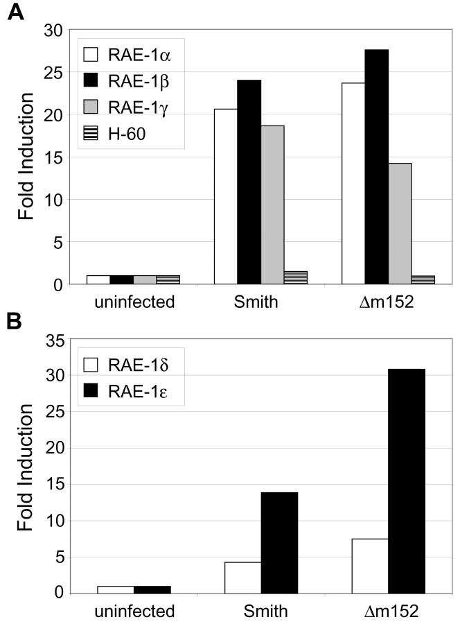
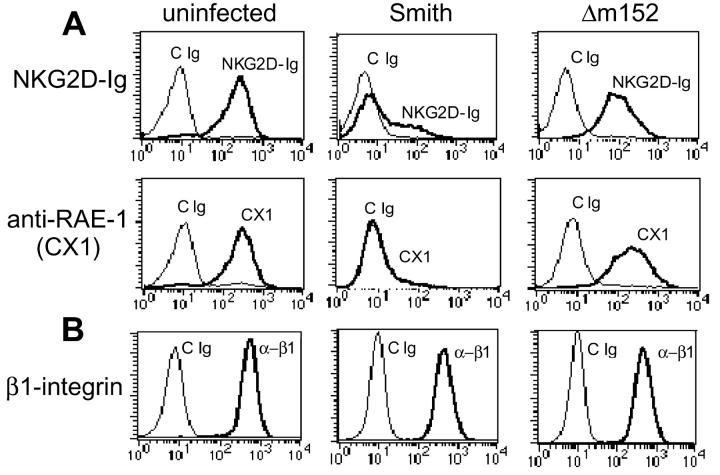
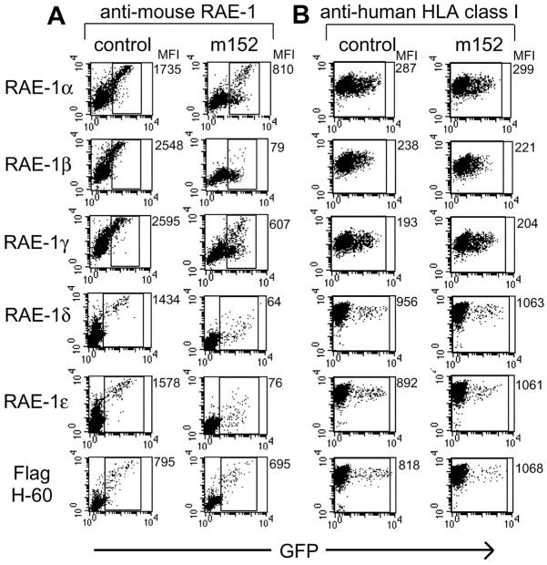
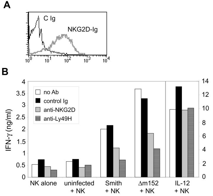
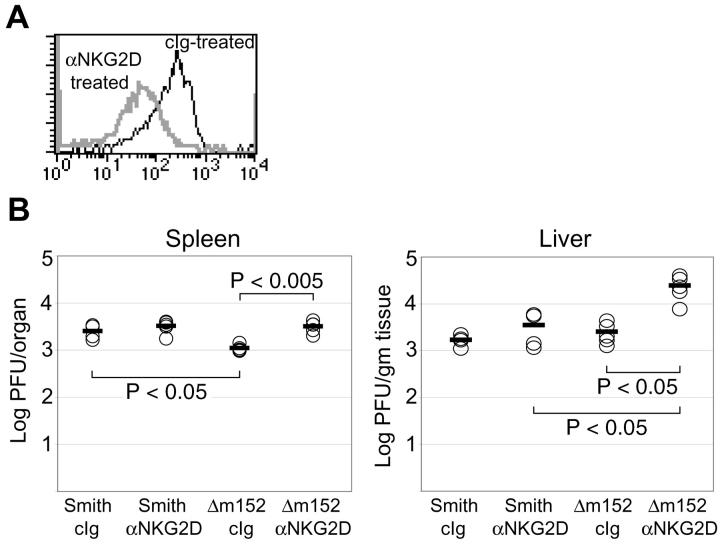
Natural killer (NK) cells play a critical role in the innate immune response against cytomegalovirus (CMV) infections. Although CMV encodes several gene products committed to evasion of adaptive immunity, viral modulation of NK cell activity is only beginning to be appreciated. A previous study demonstrated that the mouse CMV m152-encoded gp40 glycoprotein diminished expression of ligands for the activating NK cell receptor NKG2D on the surface of virus-infected cells. Here we have defined the precise ligands that are affected and have directly implicated NKG2D in immune responses to CMV infection in vitro and in vivo. Murine CMV (MCMV) infection potently induced transcription of all five known retinoic acid early inducible 1 (RAE-1) genes (RAE-1alpha, RAE-1beta, RAE-1delta, RAE-1 epsilon, and RAE-1gamma), but not H-60. gp40 specifically down-regulated the cell surface expression of all RAE-1 proteins, but not H-60, and diminished NK cell interferon gamma production against CMV-infected cells. Consistent with previous findings, a m152 deletion mutant virus (Deltam152) was less virulent in vivo than the wild-type Smith strain of MCMV. Treatment of BALB/c mice with a neutralizing anti-NKG2D antibody before infection increased titers of Deltam152 virus in the spleen and liver to levels seen with wild-type virus. These experiments demonstrate that gp40 impairs NK cell recognition of virus-infected cells through disrupting the RAE-1-NKG2D interaction.


Figures

Figure 1.
Figure 1.
RAE-1 transcripts are induced in MCMV-infected peritoneal macrophages. BALB/c and C57BL/6 peritoneal macrophages were left untreated or were infected with Smith or Δm152 (MOI = 1). RNA was extracted from cells 48 h after infection and cDNA was synthesized. TaqMan quantitative PCR was performed on BALB/c cDNA using primer/probe sets specific for RAE-1α, RAE-1β, RAE-1γ, or H-60 (A), and on C57BL/6 cDNA with primer/probe sets specific for RAE-1δ and RAE-1ɛ (B). Signals generated from each sample were normalized to HPRT signals from each set of cDNA. Data are expressed as the fold induction of transcription in infected cells compared with uninfected cells. Standard deviations of triplicate samples were <1%. Results shown are representative of three independent experiments.
Figure 2.
Figure 2.
MCMV infection results in down-regulation of NKG2D ligands. BALB/c 3T3 cells were infected with wild-type MCMV (Smith) or Δm152 (MOI = 1). 72 h after infection, infected cells were stained with control Ig (cIg), NKG2D-Ig fusion protein, or anti–RAE-1 mAb (CX1; A). As a control, cells were stained with anti–β1-integrin (B). Representative results are shown based on similar findings in three independent experiments.
Figure 3.
Figure 3.
gp40 selectively down-regulates RAE-1 proteins but not H-60. RAE-1 cDNAs or H-60 cDNA (with a 5′ Flag epitope tag) were cotransfected with a control empty vector, or with a vector encoding the m152 gene into human 293T cells. RAE-1 and H-60 cDNAs were encoded on vectors carrying an IRES-GFP whereas a non-GFP vector was used for m152 cDNA and for the control vector. 48 h after transfection, cells were stained with control Ig (cIg), biotinylated anti–RAE-1 mAb CX1 (recognizing RAE-1α, RAE-1β, and RAE-1γ), rat anti–RAE-1 mAb 186107 (recognizing RAE-1δ and RAE-1ɛ), or anti-Flag (A). As a control, cells were stained with anti–human HLA class I mAb DX17 (B). Mean fluorescence intensities (MFI) are shown to the right of each dot plot. MFI in A reflect RAE-1 or H-60 expression, calculated based on gated, GFP+ cells. MFI of human MHC class I expression (B) were determined based on the total cell population. Results are representative of similar findings in three independent experiments.
Figure 4.
Figure 4.
MCMV infection costimulates NK cell IFN-γ production through NKG2D. (A) TpnT cells were stained with a control antibody or with the NKG2D-Ig fusion protein. (B) TpnT cells were infected with Smith or Δm152 virus (MOI = 1) and cocultured with IL-2–activated C57BL/6 NK cells. Before coculture, NK cells were left untreated or treated with control Ig, anti-NKG2D (CX6), or anti-Ly49H (IF8), as indicated. As controls, NK cells were also cultured alone or in the presence of IL-12. Standard deviations of triplicate samples were <1%. This experiment was performed several times with comparable results.
Figure 5.
Figure 5.
Virulence of Δm152 virus is restored by blocking NKG2D in vivo. (A) BALB/c mice were injected intraperitoneally with control Ig or anti-NKG2D mAb (CX5). As a control to ensure NKG2D modulation, spleens were harvested from control Ig– and anti-NKG2D–treated animals 48 h after antibody treatment. Total splenocytes were stained with H-60-Ig fusion protein (to detect NKG2D), biotinylated goat anti–human Ig, streptavidin-PE, FITC-conjugated anti-pan NK cell mAb DX5 (VLA-2), and CyChrome-conjugated anti-CD3ɛ mAb. Overlaid histograms represent H-60-Ig staining of DX5+ CD3 cells from control Ig– or anti-NKG2D–treated mice. Although equivalent frequencies of DX5+ CD3 NK cells were present in control Ig– and anti-NKG2D mAb–treated mice, NKG2D was modulated on NK cells of mice treated with anti-NKG2D mAb in vivo. (B) 48 h after antibody treatment, mice were infected with 106 PFU of Smith or Δm152. 3 d after infection, mice were killed and spleens and livers were harvested. Plaque assays were performed on organ homogenates to determine viral titers in spleen and liver. Five mice were used per group. Two independent experiments were performed with comparable results; a representative experiment is shown. Statistical analysis was performed using the Student's two-tailed t test with unequal variance.

References

    1. Tortorella, D., B.E. Gewurz, M.H. Furman, D.J. Schust, and H.L. Ploegh. 2000. Viral subversion of the immune system. Annu. Rev. Immunol. 18:861–926. - PubMed
    1. Mocarski, E.S., Jr. 2002. Immunomodulation by cytomegaloviruses: manipulative strategies beyond evasion. Trends Microbiol. 10:332–339. - PubMed
    1. Kleijnen, M.F., J.B. Huppa, P. Lucin, S. Mukherjee, H. Farrell, A.E. Campbell, U.H. Koszinowski, A.B. Hill, and H.L. Ploegh. 1997. A mouse cytomegalovirus glycoprotein, gp34, forms a complex with folded class I MHC molecules in the ER which is not retained but is transported to the cell surface. EMBO J. 16:685–694. - PMC - PubMed
    1. Kavanagh, D.G., U.H. Koszinowski, and A.B. Hill. 2001. The murine cytomegalovirus immune evasion protein m4/gp34 forms biochemically distinct complexes with class I MHC at the cell surface and in a pre-Golgi compartment. J. Immunol. 167:3894–3902. - PubMed
    1. Reusch, U., W. Muranyi, P. Lucin, H.-G. Burgert, H. Hengel, and U.H. Koszinowski. 1999. A cytomegalovirus glycoprotein re-routes MHC class I complexes to lysosomes for degradation. EMBO J. 18:1081–1091. - PMC - PubMed

Publication types