. 2003 Jun 10;100(12):7259-64.
doi: 10.1073/pnas.1137463100. Epub 2003 May 19.

Involvement of multiple signaling pathways in follicular lymphoma transformation: p38-mitogen-activated protein kinase as a target for therapy

Affiliations

Involvement of multiple signaling pathways in follicular lymphoma transformation: p38-mitogen-activated protein kinase as a target for therapy

Kojo S J Elenitoba-Johnson et al. Proc Natl Acad Sci U S A. .

Abstract

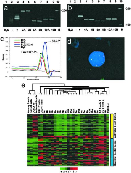
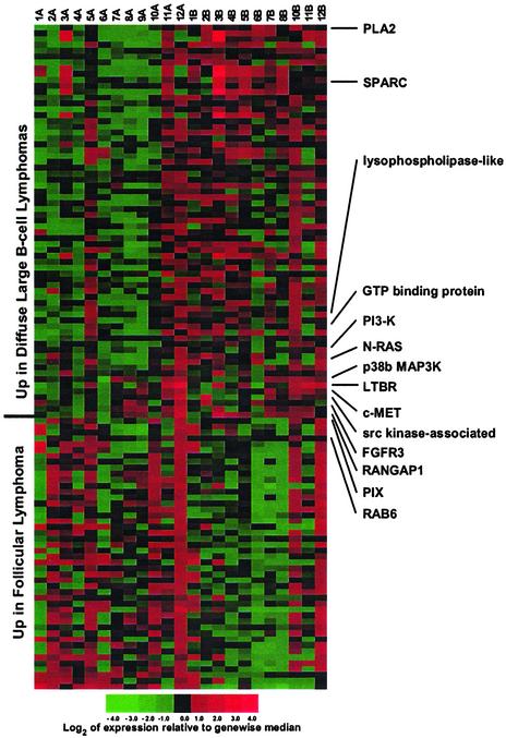
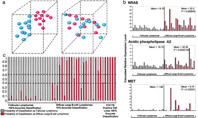
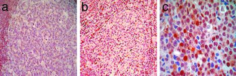
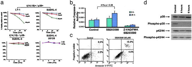
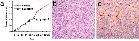
Follicular lymphoma (FL) is the most common form of low-grade non-Hodgkin's lymphoma. Transformation to diffuse large B cell lymphoma (DLBCL) is an important cause of mortality. Using cDNA microarray analysis we identified 113 transformation-associated genes whose expression differed consistently between serial clonally related samples of FL and DLBCL occurring within the same individual. Quantitative RT-PCR validated the microarray results and assigned blinded independent group of 20 FLs, 20 DLBCLs, and five transformed lymphoma-derived cell lines with 100%, 70%, and 100% accuracy, respectively. Notably, growth factor cytokine receptors and p38beta-mitogen-activated protein kinase (MAPK) were differentially expressed in the DLBCLs. Immunohistochemistry of another blinded set of samples demonstrated expression of phosphorylated p38MAPK in 6/6 DLBCLs and 1/5 FLs, but not in benign germinal centers. SB203580 an inhibitor of p38MAPK specifically induced caspase-3-mediated apoptosis in t(14;18)+/p38MAPK+-transformed FL-derived cell lines. Lymphoma growth was also inhibited in SB203580-treated NOD-SCID mice. Our results implicate p38MAPK dysregulation in FL transformation and suggest that molecular targeting of specific elements within this pathway should be explored for transformed FL therapy.


Figures

Fig. 1.
Fig. 1.
(a– d) Clonal relationship between matched pairs of FL and DLBCL. (a) IgH PCR. Lane 1, H2O control; lane 2, polyclonal control (reactive tonsil); lane 3, monoclonal control from the Raji cell line; lanes 4 and 5, monoclonal bands of identical size in the FL (2A) and the subsequent DLBCL (2B); lanes 6 and 7, monoclonal bands of identical size in the FL (8A) and DLBCL (8B) from patient 8; lanes 8 and 9, monoclonal bands of identical size in the FL (10A) and DLBCL (10B) from patient 10; and lane 10, DNA ladder. (b) bcl-2/JH (major breakpoint region, MBR) translocation. Lane 1, H2O control; lane 2, negative control (reactive tonsil); lane 3, positive control (SUDHL-6 cell line); lanes 4 and 5, bcl-2/JH products of identical size in a FL (4A) and the subsequent DLBCL (4B); lanes 6 and 7, identical bcl-2/JH product sizes in the FL and DLBCL from patient 5; lanes 8 and 9, identical bcl-2/JH product sizes in the FL (10A) and DLBCL (10B) obtained from patient 10; and lane 10, DNA ladder. (c) bcl-2(MBR)/JH product detection by fluorescence melting peak analysis. The peak at melting temperature 88.25°C represents the bcl-2/JH product from the positive control (SUDHL-6). bcl-2/JH products with a distinct melting peak with melting temperature 87.3°C is observed for both the FL (10A) and DLBCL (10B) samples from patient 10. No peak is observable in the negative control (reactive tonsil) or the H2O control. (d) t(14;18) Fluorescent in situ hybridization. One bcl-2 (green), one IgH (red), and three bcl-2/IgH (yellow) fusion signals are present. Two of the bcl-2/IgH fusion signals (yellow) represent the derivative chromosomes resulting from the reciprocal translocation. The third yellow signal represents duplication of the derivative chromosome. This fluorescent in situ hybridization pattern was detected in both the FL and DLBCL samples from patient 6. (e) Hierarchical clustering of FL and transformed FL. A total of 76 genes distinguishing GCB from the ABC-like profiles were selected from our 6,912-gene array. Clustering was performed with cluster and visualized by using tree view. Each row represents a gene and each column represents a sample. Red represents higher relative expression of a particular gene and green represents lower relative expression. The color scale at the bottom varies from -3 to +3 in log base 2 units. Consistent with their origin from GCB, the FLs and their corresponding DLBCLs exhibit a GCB-like profile.
Fig. 2.
Fig. 2.
Gene expression differences between FL and transformed FLs (DLBCLs). The FLs are designated A, and their corresponding DLBCLs are designated B. Several genes associated with MAPK signaling are differentially expressed in the transformation from FL to DLBCL.
Fig. 3.
Fig. 3.
(a) 3D separation of FL from DLBCL using three genes. The blue spheres represent the FLs and the red spheres represent their transformed counterparts (DLBCLs). (Left) Clear separation is shown in 3D using the microarray-derived expression values from PLA2 (x axis), PDGFRβ (y axis), and Rab6 (z axis). Interactive 3D animation is at www.path.utah.edu/labs/kojo. (Right) Three randomly selected genes do not separate the FL from the DLBCL. ESTs similar to KIAA0147 mRNA (x axis), human clone 23773 sequence (y axis), and diastrophic dysplasia gene (z axis). (b) Real-time PCR validation of microarray data using an independent group of FLs (n = 20) and DLBCLs (n = 20). The qRT-PCR results corroborate the results of cDNA microarray analysis and show significantly higher expression levels of N-RAS, PLA2, and C-MET in the DLBCLs. (c) Bayesian classification using a minimal discriminative gene set and qRT-PCR. Three highly ranked genes were used for classification of a blinded set of FLs and DLBCLs. The estimated probability of classification as FL (θ) ranges from 0 to 1 (gray bars). Conversely, the estimated probability of classification of the same sample as DLBCL is 1–θ (red bars). Estimated probabilities from 0.4 to 0.6 are considered indeterminate (broken horizontal lines). The accuracy rates for classification of FLs, DLBCLs, and t(14;18)-transformed lymphoma-derived cell lines were 100%, 70%, and 100%, respectively.
Fig. 4.
Fig. 4.
Expression of phosphorylated p38MAPK in transformed FL. (a) IHC demonstrates lack of phosphorylated p38MAPK expression in benign germinal centers, whereas the mantle cells demonstrate basal phosphorylated p38MAPK expression. (b) The FL also shows lack of phosphorylated p38MAPK. (c) High levels of nuclear and cytoplasmic phosphorylated p38MAPK expression are seen in the DLBCL. (Magnifications: ×40, a and b; ×400, c.)
Fig. 5.
Fig. 5.
(a) Growth inhibition of lymphoma cell lines in response to blockade of p38MAPK activity. Growth inhibition was measured by a decrease in 3-(4,5-dimethylthiazol-2-yl)-2,5-diphenyl-tetrazolium bromide conversion at 6, 24, and 48 h in comparison to DMSO-treated control cells. The results are expressed as inhibition of growth compared with control DMSO-treated cells and represent the mean ± SD of the results obtained in triplicate. Blockade of p38MAPK activity resulted in significant dose-dependent and time-dependent growth inhibition in OCI LY-1 and SUDHL-4 cells, which are t(14;18)+/p38MAPK+ (Upper). By contrast, the t(14;18)-negative/p38MAPK-negative cell line SUDHL-5 was relatively unaffected by similar doses of SB203580 (Lower Left). The specificity of p38MAPK as a target was confirmed by the lack growth inhibition by U0126 the p42/44MAPK inhibitor in the t(14;18)+/p38+ SUDHL-4 cell line (Lower Right). One representative experiment of six independent repetitions is illustrated. (b) SB203580 treatment induces caspase-3 activity. Treatment of SUDHL-4 cells with 30 μM SB203580 resulted in significant time-dependent induction of caspase-3 activity (12.6% at 6 h and 41% at 24 h; P = 0.02). Caspase-3 induction was inhibited by Z-VAD-FMK, a selective caspase-3 inhibitor. One representative experiment of three independent repetitions is illustrated. (c) Inhibition of p38MAPK induces apoptosis in t(14;18)+ cell lines. Cells were cultured in the presence of DMSO or SB203580 (50 μM) and stained for annexin V binding at the times indicated. The control represents DMSO-treated cells at 72 h. (Right) We illustrate a representative histogram for t(14;18) cell lines cultured in the presence of DMSO or SB203580 at the indicated concentrations for 72 h. Apoptosis was determined by annexin V binding and expressed as net apoptosis induction [percentage of apoptosis in treated cells minus percentage of apoptosis in control (DMSO-treated) cells]. One representative experiment of two independent repetitions is illustrated. (d) SB203580 results in selective inhibition of p38MAPK activation. Western blot analysis revealed high basal levels of phosphorylated p38MAPK in SUDHL-4 cells. Treatment with 30 μM SB203580 resulted in a significant decrease in the level of phospho-p38MAPK at 6 h. The levels of total p38MAPK and total and phospho-p42/p44 MAPK were unaffected by SB203580 treatment.
Fig. 6.
Fig. 6.
SB203580 treatment inhibits the growth of SUDHL-4 cells by apoptosis in vivo. (a) There is a significant difference in the growth of tumors in the treated versus the untreated animals. (b) In situ TUNEL assay shows only rare apoptotic (red-brown) signals in SUDHL-4 xenografts of untreated animals. (c) In situ TUNEL assay shows a significant number of apoptotic signals in comparison to untreated animals.

References

    1. Armitage, J. O. & Weisenburger, D. D. (1998) J. Clin. Oncol. 16, 2780-2795. - PubMed
    1. Horning, S. J. & Rosenberg, S. A. (1984) N. Engl. J. Med. 311, 1471-1475. - PubMed
    1. Rowley, J. D. (1988) J. Clin. Oncol. 6, 919-925. - PubMed
    1. Tsujimoto, Y., Finger, L. R., Yunis, J., Nowell, P. C. & Croce, C. M. (1984) Science 226, 1097-1099. - PubMed
    1. Hockenbery, D., Nunez, G., Milliman, C., Schreiber, R. D. & Korsmeyer, S. J. (1990) Nature 348, 334-336. - PubMed

Publication types

MeSH terms