. 2003 Jun;9(6):644-7.
doi: 10.1261/rna.5090103.

Metabolite-binding RNA domains are present in the genes of eukaryotes

Metabolite-binding RNA domains are present in the genes of eukaryotes

Narasimhan Sudarsan et al. RNA. 2003 Jun.

Abstract

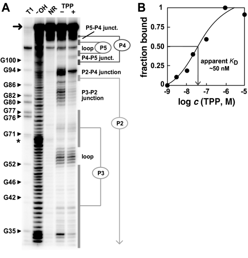
Genetic control by metabolite-binding mRNAs is widespread in prokaryotes. These riboswitches are typically located in noncoding regions of mRNA, where they selectively bind their target compound and subsequently modulate gene expression. We have identified mRNA elements in fungi and in plants that match the consensus sequence and structure of thiamine pyrophosphate-binding domains of prokaryotes. In Arabidopsis, the consensus motif resides in the 3'-UTR of a thiamine biosynthetic gene, and the isolated RNA domain binds the corresponding coenzyme in vitro. These results suggest that metabolite-binding mRNAs are possibly involved in eukaryotic gene regulation and that some riboswitches might be representatives of an ancient form of genetic control.


Figures

FIGURE 1.
FIGURE 1.
Putative eukaryote riboswitches. (A) Consensus TPP-binding domain based on 100 bacteria and archaea RNAs. Nucleotides in red are most conserved (>90%). Open circles represent nucleotide positions and domains that vary in sequence and length are designated var. The consensus model is similar to that reported recently (Rodionov et al. 2002). (B) The TPP-binding domain of A. thaliana. Variations in O. sativa (orange) and P. secunda (green) are shown. (C) Putative TPP-binding domain in the intron of N. crassa.
FIGURE 2.
FIGURE 2.
Sequence alignments of eukaryotic domains related to bacterial TPP-dependent riboswitches. Base-paired stems are shaded in black and labeled as defined previously (Winkler et al. 2002a). The P3 sequences, which in eukaryotes are significantly expanded in length and number of base pairs, are represented as a stem-loop structure. The highly conserved nucleotide positions in bacteria that were used to search for eukaryotic domains are shaded gray. For each identified (ID) sequence, the position of the conserved CUGAGA sequence within the given GenBank entry is given along with the accession identification, sequence name, and gene identification. Additional protein annotations based on sequence similarity are shown in brackets. (Methods) Riboswitch-like domains were initially identified by sequence similarity to bacterial sequences (Eco2 and Cac) by a BLASTN search of GenBank using default parameters. These hits were verified and expanded by searching for degenerate matches to the pattern (CTGAGA [200] ACYTGA [5] <<< GNTNNNNC >>> [5] CGNRGGRA) using the program SequenceSniffer (unpublished algorithm). Angle brackets indicate base pairing, and bracketed numbers are variable gaps with constrained maximum lengths. All of the eukaryotic sequences have one or zero mismatches to this pattern except for one (Aor), which initially had three mismatches due to a single A insertion in the final search element. The presence of this mutation results in an inactive aptamer, whereas removal permits TPP binding (data not shown). Comparison of mRNA (M33643.1) and genomic (AB033416.1) sequences demonstrated that the F. oxysporum element is in an intron in the 5′ UTR of the sti35 gene. Other fungal sequences (Ncr, Aor, and Fso) are flanked by consensus splicing sequences.
FIGURE 4.
FIGURE 4.
Genetic structures, thiamine biosynthetic genes, and possible mechanisms of riboswitch control. The location and mechanism of the E. coli and B. subtilis riboswitches are detailed elsewhere (Mironov et al. 2002; Winkler et al 2002a). The putative TPP riboswitch from P. secunda resides immediately upstream from the poly(A) tail in the cDNA clone of the thiC gene. The putative TPP riboswitch domain in F. oxysporum is located in a 5′-UTR intron of the sti35 gene according to the genomic sequence, but is absent in the cDNA clone.
FIGURE 3.
FIGURE 3.
Structural probing of the putative TPP-riboswitch from Arabidopsis. (A) Fragmentation pattern of the 128-nucleotide RNA (arrow) of A. thaliana (Fig. 1B ▶), which was generated by incubation in the absence (−) or presence (+) of 100 μM TPP. T1, OH, and NR identify RNAs that were partially digested with RNase T1 (cleaves 3′ to G residues), alkali, or were not reacted, respectively. Reactions were conducted as described previously (Winkler et al. 2002a). (B) Apparent KD for TPP binding by the A. thaliana RNA. Fraction bound was determined by in-line probing as described previously (Nahvi et al. 2002; Winkler et al. 2002a,b).

Similar articles

Cited by

References

    1. Begley, T.P., Downs, D.M., Ealick, S.E., McLafferty, F.W., Van Loon, A.P., Tayla, S., Campobasso, N., Chiu, H.J., Kinsland, C., Reddick, J.J., et al. 1999. Thiamin biosynthesis in prokaryotes. Arch. Microbiol. 171: 293–300. - PubMed
    1. Benner, S.A., Ellington, A.D., and Tauer, A. 1989. Modern metabolism as a palimpsest of the RNA world. Proc. Natl. Acad. Sci. 86: 7054–7058. - PMC - PubMed
    1. Gelfand, M.S., Mironov, A.A., Jomantas, J., Kozlov, Y.I., and Perumov, D.A. 1999. A conserved RNA structure element involved in the regulation of bacterial riboflavin synthesis genes. Trends Genet. 15: 439–442. - PubMed
    1. Hesselberth, J.R., Robertson, M.P., Knudsen, S.M., and Ellington, A.D. 2003. Simultaneous detection of diverse analytes with aptazyme ligase array. Anal. Biochem. 312: 106–112. - PubMed
    1. Jeffares, D.C., Poole, A.M., and Penny, D. 1998. Relics from the RNA world. J. Mol. Evol. 46: 18–36. - PubMed

Publication types