. 2003 Jun;9(6):677-87.
doi: 10.1261/rna.2128903.

An uncapped RNA suggests a model for Caenorhabditis elegans polycistronic pre-mRNA processing

Affiliations

An uncapped RNA suggests a model for Caenorhabditis elegans polycistronic pre-mRNA processing

Yingmiao Liu et al. RNA. 2003 Jun.

Abstract

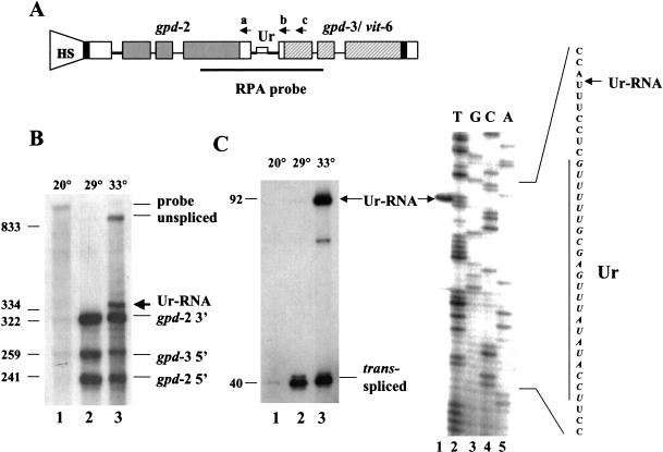
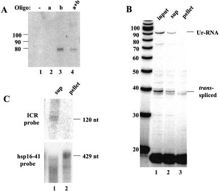
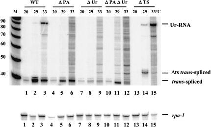
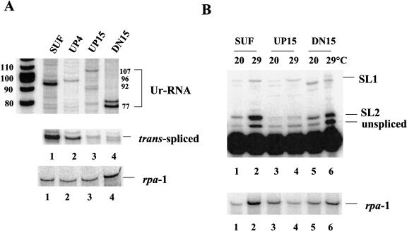
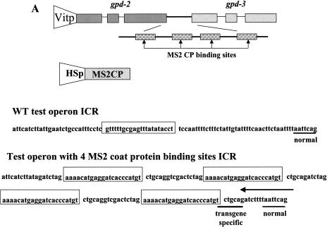
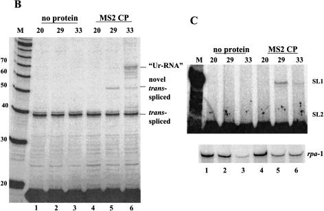
Polycistronic pre-mRNAs from Caenohabditis elegans operons are processed by internal cleavage and polyadenylation to create 3' ends of mature mRNAs. This is accompanied by trans-splicing with SL2 approximately 100 nucleotides downstream of the 3' end formation sites to create the 5' ends of downstream mRNAs. SL2 trans-splicing depends on a U-rich element (Ur), located approximately 70 nucleotides upstream of the trans-splice site in the intercistronic region (ICR), as well as a functional 3' end formation signal. Here we report the existence of a novel gene-length RNA, the Ur-RNA, starting just upstream of the Ur element. The expression of Ur-RNA is dependent on 3' end formation as well as on the presence of the Ur element, but does not require a trans-splice site. The Ur-RNA is not capped, and alteration of the location of the Ur element in either the 5' or 3' direction alters the location of the 5' end of the Ur-RNA. We propose that a 5' to 3' exonuclease degrades the precursor RNA following cleavage at the poly(A) site, stopping when it reaches the Ur element, presumably attributable to a bound protein. Part of the function of this protein can be performed by the MS2 coat protein. Recruitment of coat protein to the ICR in the absence of the Ur element results in accumulation of an RNA equivalent to Ur-RNA, and restores trans-splicing. Only SL1, however, is used. Therefore, coat protein is sufficient for blocking the exonuclease and thereby allowing formation of a substrate for trans-splicing, but it lacks the ability to recruit the SL2 snRNP. Our results also demonstrate that MS2 coat protein can be used as an in vivo block to an exonuclease, which should have utility in mRNA stability studies.


Figures

FIGURE 1.
FIGURE 1.
High-temperature heat shock results in accumulation of a novel RNA containing part of the intercistronic region. (A) The HS1496 construct. The position of the heat shock promoter is shown by the shape marked HS. (Shaded bars) Coding regions of gpd-2; (hatched bars) coding regions of gpd-3; (solid bar) coding region of vit-6; (open bars) noncoding regions of exons; (thin lines) introns; (thicker lines) intercistronic sequences. The Ur element is indicated by a narrow bar. The dark line below the operon diagram indicates the region covered by the RNase protection probe. Antisense oligonucleotides a, b, and c are indicated above the positions to which they can anneal. (B) RNase protection assay of total RNA from the transgenic worm strain BL4057, carrying the HS1496 construct. Worms were grown at 20°C and heat shocked at 29°C or 33°C for 2 h. RNA was isolated from 20°C controls as well as the two heat-shocked populations for RPA. The sizes and identifications of the protected fragments were calculated and indicated. Low levels of products seen in lane 1 represent endogenous gpd-2 and gpd-3 transcripts. (C) Primer extension analysis of RNA preparations analyzed in B. Ur-RNA and trans-spliced products are indicated. In the gel at right, location of the 5′ end of Ur-RNA is determined. The gpd-2/gpd-3 intercistronic region was sequenced using the same oligonucleotide used for primer extension and compared with the primer extension product as in the gel shown on the left (lane 3). Part of the intercistronic region sequence is depicted on the right with the Ur element indicated by a vertical line.
FIGURE 2.
FIGURE 2.
Ur-RNA is monocistronic and uncapped. (A) Northern analysis of RNase H digested Ur-RNA. BL4057 RNA (33°C) was annealed with oligonucleotides a, b, or both (Fig. 1A ▶), treated with RNase H, separated on an 8% denaturing polyacrylamide gel and blotted. The blot was probed with a random-primed probe against the intercistronic region (ICR probe). The band just below the 80-nucleotide marker is the size predicted for an RNA beginning at the 5′ end of Ur-RNA and ending at the b oligonucleotide. (B) Fractionation of capped and uncapped RNA on eIF4E resin. BL4057 RNA (33°C) was annealed with oligonucloetide c, treated with RNase H, and affinity purified with GST–eIF4E fusion protein. RNAs from both supernatant and pellet were analyzed by primer extension. (C) Northern blot analysis of the RNase H-digested, eIF4E-fractionated RNA, probed with the ICR probe and with hsp-16-41 probe as control.
FIGURE 3.
FIGURE 3.
Both the Ur element and the poly (A) signal are required for Ur-RNA formation. Total RNA from transgenic strains carrying HS1496 (WT) and various mutations of the HS1496 construct were analyzed by primer extension. Ur-RNA and trans-spliced products are indicated. The band above the trans-spliced product that appears in lane 14 is attributable to usage of a cryptic trans-splice site (see text). Similar amounts of RNA were subjected to primer extension in each lane as indicated by the rpa-1 primer extension product (lower panel).
FIGURE 4.
FIGURE 4.
The location of the Ur element determines the location of the 5′ end of Ur-RNA and influences the efficiency of trans-splicing. (A) Transgenic worm strains were grown and heat shocked at 33°C for 2 h. RNA was isolated and analyzed by primer extension. Primer extension was performed on RNA isolated from several strains carrying each construct, but data from only a single representative strain is shown. The predicted sizes of Ur-RNAs from the four constructs are indicated on the right. The trans-spliced products are shown in the lower panel. (B) RNA from strains grown at 20°C and heat shocked at 29°C was subjected to primer extension in the presence of ddGTP. Positions expected for unspliced precursor, SL2, and SL1 trans-spliced products (2-, 3-, and 9-nucleotide extensions, respectively) are shown on the right. Rpa-1 primer extension in the lower panel of both A and B serves as a loading control.
FIGURE 5.
FIGURE 5.
MS2 coat protein bound to the ICR promotes SL1 trans-splicing. (A) The MS2 constructs. The test operon construct is composed of the gpd-2/gpd-3 operon under control of the vit-2 promoter, with the gpd-2/gpd-3 intercistronic sequence replaced by an ICR containing four MS2 coat protein-binding sites. Exons, introns, and the ICR are shown to scale. The ICR containing the coat protein-binding sites (to scale) is enlarged for clarity. The open reading frame of MS2 coat protein is under control of the hsp-16-41 promoter. The ICR sequence is shown for both the gpd-2/gpd-3 operon and the MS2 construct, with the Ur element and the MS2 coat protein binding sites boxed. The normal and transgene-specific trans-splice sites are underscored. The arrow above the sequence indicates the 3′ end of the primer that targets only the transgene. (B) Primer extension with the primer that spans the 5′ end of gpd-3. “Ur-RNA” and trans-spliced products are indicated. (Lanes 1–3) No coat protein transgene present; (lanes 4–6) with coat protein transgene. (Lanes 1,4) not heat shocked; (lanes 2,3,5,6) heat shocked at the temperatures indicated above the lanes. (M) Markers. (C) Primer extension with the transgene-specific primer in the presence of ddGTP. Positions expected for SL1 and SL2 trans-spliced products are shown on the right. Only SL1 trans-spliced products are observed. Lane designations are as in B. The lower panel shows rpa-1 primer extension as a loading control.
FIGURE 5.
FIGURE 5.
MS2 coat protein bound to the ICR promotes SL1 trans-splicing. (A) The MS2 constructs. The test operon construct is composed of the gpd-2/gpd-3 operon under control of the vit-2 promoter, with the gpd-2/gpd-3 intercistronic sequence replaced by an ICR containing four MS2 coat protein-binding sites. Exons, introns, and the ICR are shown to scale. The ICR containing the coat protein-binding sites (to scale) is enlarged for clarity. The open reading frame of MS2 coat protein is under control of the hsp-16-41 promoter. The ICR sequence is shown for both the gpd-2/gpd-3 operon and the MS2 construct, with the Ur element and the MS2 coat protein binding sites boxed. The normal and transgene-specific trans-splice sites are underscored. The arrow above the sequence indicates the 3′ end of the primer that targets only the transgene. (B) Primer extension with the primer that spans the 5′ end of gpd-3. “Ur-RNA” and trans-spliced products are indicated. (Lanes 1–3) No coat protein transgene present; (lanes 4–6) with coat protein transgene. (Lanes 1,4) not heat shocked; (lanes 2,3,5,6) heat shocked at the temperatures indicated above the lanes. (M) Markers. (C) Primer extension with the transgene-specific primer in the presence of ddGTP. Positions expected for SL1 and SL2 trans-spliced products are shown on the right. Only SL1 trans-spliced products are observed. Lane designations are as in B. The lower panel shows rpa-1 primer extension as a loading control.

Similar articles

Cited by

References

    1. Blumenthal, T., Evans, D., Link, C.D., Guffanti, A., Lawson, D., Thierry-Mieg, J., Thierry-Mieg, D., Chiu, W.L., Duke, K., Kiraly, M., et al. 2002. A global analysis of Caenorhabditis elegans operons. Nature 417: 851–854. - PubMed
    1. Blumenthal, T. and Spieth, J. 1996. Gene structure and organization in Caenorhabditis elegans. Curr. Opin. Genet. Dev. 6: 692–698. - PubMed
    1. Brenner, S. 1974. The genetics of Caenorhabditis elegans. Genetics 77: 71–94. - PMC - PubMed
    1. Conrad, R., Thomas, J., Spieth, J., and Blumenthal, T. 1991. Insertion of part of an intron into the 5′ untranslated region of a Caenorhabditis elegans gene converts it into a trans-spliced gene. Mol. Cell. Biol. 11: 1921–1926. - PMC - PubMed
    1. Conrad, R., Liou, R.F., and Blumenthal, T. 1993. Conversion of a trans-spliced C. elegans gene into a conventional gene by introduction of a splice donor site. EMBO J. 12: 1249–1255. - PMC - PubMed

Publication types

MeSH terms

LinkOut - more resources