. 2003 Jun;9(6):688-97.
doi: 10.1261/rna.2600503.

Substrate specificity and reaction kinetics of an X-motif ribozyme

Affiliations

Substrate specificity and reaction kinetics of an X-motif ribozyme

Denis Lazarev et al. RNA. 2003 Jun.

Abstract

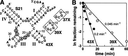
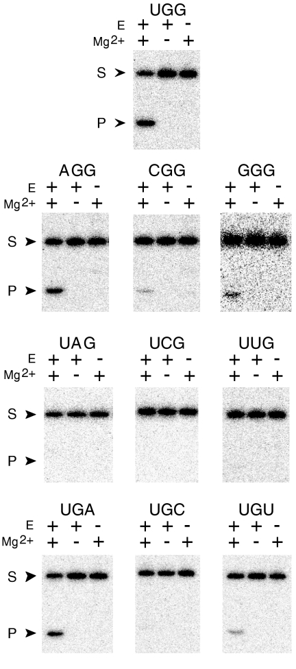
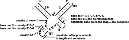
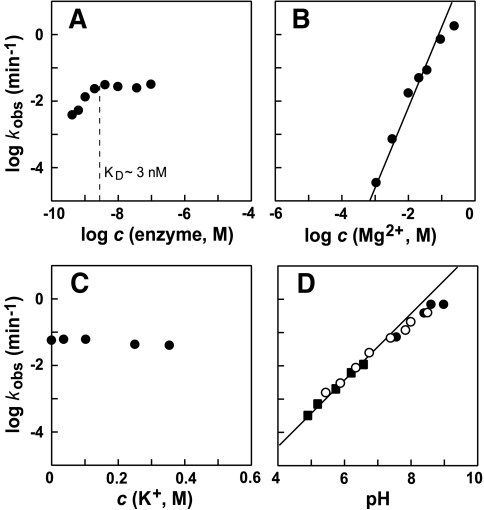
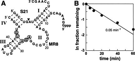
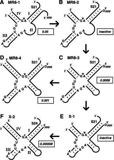
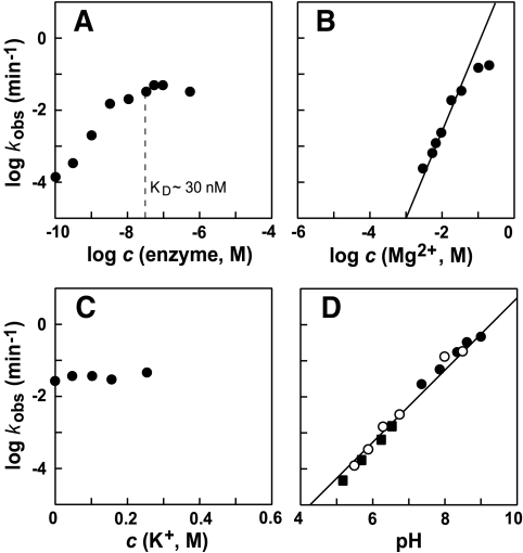
The X-motif is an in vitro-selected ribozyme that catalyzes RNA cleavage by an internal phosphoester transfer reaction. This ribozyme class is distinguished by the fact that it emerged as the dominant clone among at least 12 different classes of ribozymes when in vitro selection was conducted to favor the isolation of high-speed catalysts. We have examined the structural and kinetic properties of the X-motif in order to provide a framework for its application as an RNA-cleaving agent and to explore how this ribozyme catalyzes phosphoester transfer with a predicted rate constant that is similar to those exhibited by the four natural self-cleaving ribozymes. The secondary structure of the X-motif includes four stem elements that form a central unpaired junction. In a bimolecular format, two of these base-paired arms define the substrate specificity of the ribozyme and can be changed to target different RNAs for cleavage. The requirements for nucleotide identity at the cleavage site are GD, where D = G, A, or U and cleavage occurs between the two nucleotides. The ribozyme has an absolute requirement for a divalent cation cofactor and exhibits kinetic behavior that is consistent with the obligate binding of at least two metal ions.


Figures

FIGURE 1.
FIGURE 1.
Bimolecular constructs of X-motif ribozymes. (A) Sequence and secondary-structure model of 43X, 39X, and 37X in complex with the S21 substrate RNA. Roman numerals I through IV identify four putative stems that are characteristic of X-motif ribozymes. Encircled nucleotides are conserved in the ribozyme portion of all X-motif variants encountered thus far. The arrowhead identifies the site of ribozyme-catalyzed RNA cleavage. The lower and upper insets depict the shortened stem II elements that are present in 39X and 37X, respectively. (B) Determination of rate constants for S21 cleavage by 43X and 39X ribozymes. The fraction of S21 cleaved in the presence of 43X (filled circles) versus 39X (open circles) is plotted relative to incubation time, after correction for the amount of substrate that remains uncleaved on extended incubation (see Materials and Methods). Reactions contained 50 mM Tris-HCl (pH 7.5 at 23°C), 250 mM KCl, 20 mM MgCl2, trace amounts of 5′ 32P-labeled S21, and 50 nM of 43X or 39X RNAs. The concentration of 43X and 39X used ensures that all substrate is bound to ribozyme. The kobs value for each ribozyme was established by determining the negative slope of the corresponding line.
FIGURE 2.
FIGURE 2.
Sequence requirements for an X-motif ribozyme. The trinucleotide depicted above each panel designates the sequence of nucleotides at positions 8, 9, and 10 of each S21 substrate variant tested. The original substrate carries a UGG sequence at these positions, and the activity of the 39X ribozyme toward this substrate is depicted in the top panel. The activity of this ribozyme toward all possible one-nucleotide base changes relative to the UGG substrate is depicted in subsequent panels. Each assay contained a trace amount of 5′ 32P-labeled S21 or similarly labeled variant S21 RNA, 50 mM Tris-HCl (pH 7.5 at 23°C), and 250 mM KCl. The reactions were incubated at 23°C for 15 min in the absence (−) or presence (+) of 50 nM 39X (E) and 20 mM MgCl2 (Mg2+), as indicated. The uncleaved substrate (S) and 5′-cleavage product (P) were separated by denaturing 20% PAGE, and an autoradiogram is depicted for each analysis.
FIGURE 3.
FIGURE 3.
Important sequence and structural elements of X-motif ribozymes. Stems I through IV are supported by artificial phylogeny data, or have been confirmed by mutational analysis. Letters identify strictly conserved nucleotides, numbers identify conserved base-pairing interactions, and filled circles identify positions that favor specific nucleotide identities, as denoted. The arrowhead indicates the site of cleavage.
FIGURE 4.
FIGURE 4.
Kinetic characteristics of S21 cleavage by the 39X ribozyme. In each graph, the logarithm of the rate constant for RNA cleavage is plotted relative to the concentration of the various agents, as indicated. (A) Dependence of the rate constant on the concentration of ribozyme. Reactions contained trace amounts of 5′ 32P-labeled S21, 50 mM Tris-HC1 (pH 7.5 at 23°C), 250 mM KCl, and 20 mM MgCl2. Rate constants were determined as described in Materials and Methods. (B) Dependence of rate constant on the concentration of divalent magnesium. Reactions were conducted as described in A, but the concentration of 39X was held constant at 50 nM, and the concentration of MgCl2 was varied, as indicated. The line represents a slope of 2. (C) Dependence of rate constant on the concentration of potassium ions. Reactions were conducted as described in B, but the concentration of MgCl2 was held constant at 20 mM, and the concentration of KCl was varied, as indicated. (D) Dependence of rate constant on pH. Reactions were conducted as described in C, but KCl was omitted from the reaction, and the pH was varied, as indicated. Filled circles, open circles, and filled squares identify data points generated with reactions that were buffered with Tris, HEPES, and MES, respectively. The line represents a slope of 1.
FIGURE 5.
FIGURE 5.
The structure and function of the MR8–1 ribozyme. (A) Sequence and secondary structure model of the MR8–1 ribozyme in complex with S21 RNA substrate. Roman numerals I through IV identify stem elements and an unstructured region (II) that correspond either in structure or in location to the four stems of X-motif ribozymes. The arrowhead identifies the site of ribozyme cleavage and encircled nucleotides match the conserved core of the X-motif ribozymes. (B) Determination of rate constant for S21 cleavage by MR8–1. The analysis was conducted as described in the legend to Figure 1B ▶.
FIGURE 6.
FIGURE 6.
Constructs used to explore the similarity between the MR8 and X-motif classes of ribozymes. (A) The MR8–1 construct is identical to that of the MR8 sequence depicted in Figure 5A ▶, with the exception of a single G-to-A change at position 11 that was made to establish complete Watson/Crick base pairing in stem I. (B) Construct MR8–2 is identical to MR8–1, wherein the 5′-most nucleotides are replaced by a single G residue. (C) Construct MR8–3 is identical to MR8–2, wherein the 11-nucleotide internal bulge (II) is replaced by a U residue at position 14 and a 12-nucleotide stem-loop sequence that is identical to stem II of the 43X ribozyme depicted in Figure 1A ▶. (D) Construct MR8–4 is identical to MR8–3, wherein the substrate base-pairing residues of stems I and IV are altered to target S21 cleavage between nucleotides 9 and 10 (the original X-motif cleavage site). (E) Construct X-1 is identical to 39X (Fig. 1A ▶, inset), wherein the substrate base-pairing residues of stems I and IV are altered to target S21 cleavage between nucleotides 6 and 7 (the original MR8 cleavage site). (F) Construct X-2 is identical to construct X-1, wherein the base-pairing region of stem IV has been extended by three nucleotides to create eight base pairs with the variant substrate molecule S24. S24 is identical in sequence to S21, except that three nucleotides (5′-GUC) have been added to the 5′ terminus. Boxed numbers represent the rate constants that were measured (MR8–1 and X-2) or that were estimated based on single-time-point assays. Rate constants (min−1) are boxed. Constructs with undetectable levels of RNA cleavage (k values below 1 × 10⁻⁵ min−1) are identified as inactive.
FIGURE 7.
FIGURE 7.
Kinetic characteristics of S21 cleavage by the MR8–1 ribozyme. Details are as described in the legend to Figure 4 ▶ except that 250 mM KCl was present in assays that provided data for D.

Similar articles

Cited by

References

    1. Doherty, E.A. and Doudna, J.A. 2000. Ribozyme structure and mechanisms. Annu. Rev. Biochem. 69: 597–615. - PubMed
    1. Forster, A.C. and Symons, R.H. 1987. Self-cleavage of plus and minus RNAs of a virusoid and a structural model for the active sites. Cell 49: 211–220. - PubMed
    1. Hertel, K.J., Pardi, A., Uhlenbeck, O.C., Koizumi, M., Ohtsuka, E., Uesugi, S., Cedergren, R., Eckstein, F., Gerlach, W.L., Hodgson, R., et al. 1992. Numbering system for the hammerhead. Nucleic Acids Res. 20: 3252. - PMC - PubMed
    1. Ishizaka, M., Oshima, Y., and Tani, T. 1995. Isolation of active ribozymes from an RNA pool of random sequences using an anchored substrate RNA. Biochem. Biophys. Res. Commun. 214: 403–409. - PubMed
    1. James, H.A. and Gibson, I. 1998. The therapeutic potential of ribozymes. Blood 91: 371–382. - PubMed

Publication types