

Isolation of proliferation factor of immature T-cell clone in concanavalin A-stimulated splenocyte culture supernatant
- PMID: 12757615
- PMCID: PMC1782958
- DOI: 10.1046/j.1365-2567.2003.01642.x
Isolation of proliferation factor of immature T-cell clone in concanavalin A-stimulated splenocyte culture supernatant
Abstract
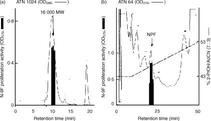
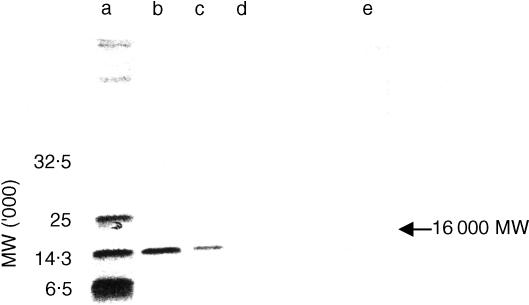
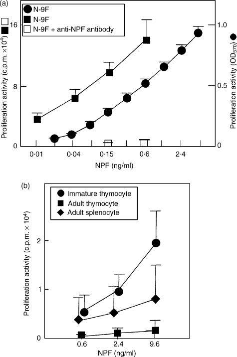
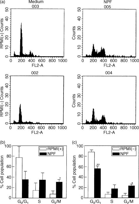
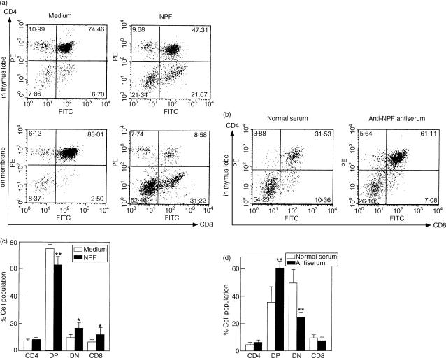
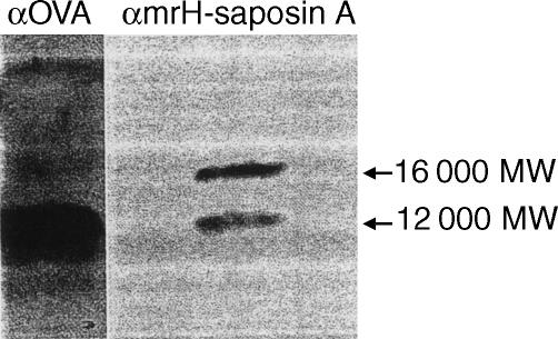
An athymic mouse-derived immature T-cell clone, N-9F, was not maintained by interleukin-2 alone but required another soluble factor, contained in concanavalin A-stimulated rat splenocyte culture supernatant, namely T cell growth factor (TCGF), for its proliferation. An N-9F-proliferation factor (NPF) was isolated in a pure form from TCGF. N-9F cells and immature thymocytes proliferated in the presence of NPF at 10-11-10-8 g/ml in a dose-dependent manner, but adult thymocytes were not stimulated by NPF. NPF increased DNA synthesis of N-9F. NPF increased CD4 and CD8 double negative thymocytes and CD8 single positive thymocytes in fetal thymus organ culture. A hamster anti-NPF antiserum possessing the capacity to neutralize N-9F proliferation activity of NPF decreased double negative thymocytes. The amino-terminal amino acid sequence of NPF was identified to be Ser-Leu-Pro-Cys-Asp-Ile-Cys-Lys-Thr-Val-Val-Thr-Glu-Ala-Cys-Asn-Leu-Leu-Lys-Asp- and was identical to that of rat saposin A. The apparent molecular weight of NPF, 16000, was comparable to that of saposin A. A rabbit anti-mouse recombinant His-tag (mrH)-saposin A antibody recognized a 16000 MW molecule in TCGF. A Hitrap-saposin A antibody column bound NPF and pulled down the NPF activity in TCGF. Thus, NPF in TCGF was a saposin A-like protein possessing the capacity for growth and differentiation of immature thymocytes.
Figures
Similar articles
-
[Discovery of immature thymocyte proliferation factor].Yakugaku Zasshi. 2006 Mar;126(3):145-60. doi: 10.1248/yakushi.126.145. Yakugaku Zasshi. 2006. PMID: 16508238 Review. Japanese.
-
Proliferation of an athymic mouse-derived T-cell clone on thymic stromal cells with interleukin-2.Immunology. 1991 Oct;74(2):264-70. Immunology. 1991. PMID: 1748473 Free PMC article.
-
IL-10, a novel growth cofactor for mature and immature T cells.J Immunol. 1990 Dec 15;145(12):4167-73. J Immunol. 1990. PMID: 2124236
-
Proliferation and differentiation of single hapten-specific B lymphocytes is promoted by T-cell factor(s) distinct from T-cell growth factor.Proc Natl Acad Sci U S A. 1982 Oct;79(20):6350-4. doi: 10.1073/pnas.79.20.6350. Proc Natl Acad Sci U S A. 1982. PMID: 6983069 Free PMC article.
-
Thymocyte growth factor: a progression growth factor for cycling immature cortical thymocytes.Comp Immunol Microbiol Infect Dis. 1985;8(3-4):235-46. doi: 10.1016/0147-9571(85)90002-5. Comp Immunol Microbiol Infect Dis. 1985. PMID: 3912098 Review.
References
-
- Von Boehmer H. The developmental biology of T lymphocytes. Annu Rev Immunol. 1987;6:309–26. - PubMed
-
- Haynes BF, Markert ML, Sempowski GD, Patel DD, Hale LP. The role of the thymus in immune reconstitution in aging, bone marrow transplantation, and HIV-1 infection. Annu Rev Immunol. 2000;18:529–60. - PubMed
-
- Shortman K, Wu L. Early thymocyte progenitors. Annu Rev Immunol. 1996;14:29–47. - PubMed
-
- Anderson G, Moore NC, Owen JJT, Jenkinson EJ. Cellular interactions in thymocyte development. Annu Rev Immunol. 1996;14:73–99. - PubMed
-
- Ardavin C. Thymic dendritic cells. Immunol Today. 1997;18:350–61. - PubMed
Publication types
MeSH terms
Substances
LinkOut - more resources
Full Text Sources
Research Materials
