. 2003 Jun;109(2):226-31.
doi: 10.1046/j.1365-2567.2003.01648.x.

Costimulation through OX40 is crucial for induction of an alloreactive human T-cell response

Affiliations

Costimulation through OX40 is crucial for induction of an alloreactive human T-cell response

Naoya Ukyo et al. Immunology. 2003 Jun.

Abstract

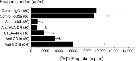
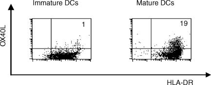
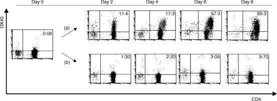
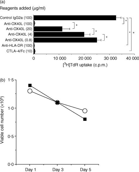
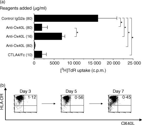
The alloreactive immune response is a series of events initiated by the interaction of T cells with allogeneic dendritic cells (DCs), involving alloantigen recognition and costimulatory signals. In this study, we investigated the role of OX40 in alloreactivity in vitro. We first demonstrated that anti-OX40 ligand (anti-OX40L) monoclonal antibody (mAb) could markedly suppress the mixed leucocyte reaction (MLR) of peripheral blood mononuclear cells (PBMC). To further define the contribution of the OX40/OX40L system to the MLR, we set up a co-culture system of CD4+ T cells and allogeneic monocyte-derived dendritic cells (DCs). After 2 days, OX40 expression was induced on CD4+ T cells and this induction was strongly inhibited by the addition of cytotoxic T lymphocyte-associated antigen-4 (CTLA-4)-Fc fusion protein, suggesting that the expression of OX40 during alloreaction is dependent on CD28 signalling. Next we examined the effects of anti-OX40L mAb, CTLA-4-Fc fusion protein and anti-human leucocyte antigen (HLA)-DR mAb on the proliferative response of CD4+ T cells to allogeneic DCs. The proliferation of T cells was almost completely suppressed by anti-OX40L mAb, which was comparable with that of CTLA-4-Fc. Measurement of interleukin-2 (IL-2) production in the culture supernatants showed that suppression of a proliferative response was at least in part ascribed to reduced IL-2 production. Furthermore, purified OX40L- allogeneic DCs could induce considerable proliferation of CD4+ T cells, which was suppressed by anti-OX40L mAb. These results suggest that the OX40/OX40L system is crucial for induction of the allogeneic T-cell response and the OX40/OX40L system is subsequent to and dependent on CD28 signalling, but is crucial for the end outcome of the human alloreactive T-cell response.


Figures

Figure 1
Figure 1
Effects of anti-OX40 ligand (OX40L), anti-human leucocyte antigen (HLA)-DR, anti-CD18 and cytotoxic T lymphocyte-associated antigen-4 (CTLA-4)-Fc fusion protein on the two-way mixed leucocyte reaction (MLR). Peripheral blood mononuclear cells (PBMC) from two different donors were mixed and co-cultured in a 96-well culture plate for 7 days in the presence of the indicated reagents. Cells were pulsed with [3H]thymidine ([3H]TdR) (0·5 µCi/well) for the last 7 hr of culture and the incorporated radioactivity was measured in a liquid scintillation counter. Results are expressed as mean counts ± standard deviation (SD) of triplicate samples. The asterisk indicates a P-value of < 0·05 compared with the suppressed reactivity.
Figure 2
Figure 2
OX40 ligand (OX40L) expression on monocyte-derived dendritic cells (DC), in the presence or absence of stimulation with monocyte-conditioned medium (MCM). Purified monocytes were cultured in RPMI-1640 supplemented with granulocyte–macrophage colony-stimulating factor (GM-CSF), interleukin-4 (IL-4) and autologous plasma for 7 days to generate immature DCs. Then, cells were cultured for a further 2 days, with or without MCM. Cells were stained with fluorescein isothiocyanate (FITC)-conjugated anti-human leucocyte antigen (HLA)-DR, biotinylated anti-OX40L (ik-1), and phycoerythrin (PE)-labelled streptavidin, and subsequent flow cytometric analysis, with a FACScan, was performed. The percentage of OX40L+ HLA-DR+ cells is indicated in the upper right corner of each dot-plot.
Figure 3
Figure 3
OX40 expression on CD4+ T cells stimulated by allogeneic dendritic cells (DCs) and the inhibitory effect of cytotoxic T lymphocyte-associated antigen-4 (CTLA-4)-Fc fusion protein against the expression of OX40. CD4+ T cells and allogeneic irradiated mature DCs (mDCs) were co-cultured in a 96-well culture plate at a ratio of 200 : 1. (a) Control monoclonal antibody (mAb) (MOPC21) or (b) CTLA-4–Fc fusion protein was added to the culture at a final concentration of 100 or 10 µg/ml, respectively. On days 0, 2, 4, 6 and 8, cells were collected, stained with fluorescein isothiocyanate (FITC)-conjugated anti-CD4, biotinylated anti-OX40L (ik-1) and phycoerythrin (PE)-labelled streptavidin, and subjected to flow cytometric analysis in a FACScan. The percentage of OX40+ CD4+ T cells is indicated in the upper right corner of each dot-plot.
Figure 4
Figure 4
Effects of anti-OX40 ligand (OX40L) monoclonal antibody (mAb) on CD4+ T-cell proliferation stimulated by allogeneic dendritic cells (DCs). (a) CD4+ T cells and allogeneic irradiated mature DCs (mDCs) were mixed and co-cultured in a 96-well culture plate for 5 days in the presence of the indicated reagents. Cells were pulsed with [3H]thymidine ([3H]TdR) (0·5 µCi/well) for the last 7 hr of culture and the incorporated radioactivity was measured in a liquid scintillation counter. Results are expressed as mean counts ± standard deviation (SD) of triplicate samples. The asterisk indicates a P-value of < 0·05 compared with the suppressed reactivity. (b) Effects of anti-OX40L mAb on the viability of DCs. DCs were cultured in RPMI-1640 supplemented with 10% fetal calf serum (FCS) in the presence of 100 µg/ml control mouse IgG2a (UPC-10; open circle) or anti-OX40L mAb (ik-5; closed square) and the viable cell number was counted by using Trypan-Blue dye exclusion.
Figure 5
Figure 5
Effects of anti-OX40 ligand (OX40L) monoclonal antibody (mAb) on the proliferation of CD4+ T cells stimulated by purified OX40L mature DCs (mDCs). (a) OX40L mDCs were purified in a cell sorter and irradiated. CD4+ T cells and OX40L mDCs were mixed at a ratio of 1000 : 1 and co-cultured in a 96-well culture plate for 7 days in the presence of the indicated reagents. Cells were pulsed with [3H]thymidine ([3H]TdR) (0·5 µCi/well) for the last 7 hr of culture and the incorporated radioactivity was measured in a liquid scintillation counter. Results are expressed as mean counts ± standard deviation (SD) of triplicate samples. The asterisk indicates a P-value of < 0·05 for comparison with the suppressed reactivity. (b) CD4+ T cells and allogeneic OX40L mDCs were mixed at a ratio of 10 : 1 and co-cultured in a 96-well culture plate for 7 days. Cells were collected on days 3, 5 and 7, and the expression of OX40L was examined on mDCs.

Similar articles

Cited by

References

    1. Steinmann RM, Gutchinov B, Witmer MD, Nussenzweig MC. Dendritic cells are the principal stimulators of the primary mixed leukocyte reaction in mice. J Exp Med. 1983;157:613–27. - PMC - PubMed
    1. Caux C, Vanbervliet B, Massacrier C, Azuma M, Okumura K, Lanier LL, Banchereau J. B70/B7-2 is identical to CD86 and is the major functional ligand for CD28 expressed on human dendritic cells. J Exp Med. 1994;180:1841–7. - PMC - PubMed
    1. Tanaka H, Demeure CE, Rubio M, Delespesse G, Saefati M. Human monocyte-derived dendritic cells induce naive T cell differentiation into T helper cell type 2 (Th2) or Th1/Th2 effectors: role of stimulator/responder ratio. J Exp Med. 2000;192:405–12. - PMC - PubMed
    1. Ferlazzo G, Wesa A, Wei WZ, Galy A. Dendritic cells generated either from CD34+ progenitor cells or monocytes differ in their ability to activate antigen-specific CD8+ T cells. J Immunol. 1999;163:3597–604. - PubMed
    1. Aicher A, Hayden-Ledbetter M, Brady WA, et al. Characterization of human inducible costimulator ligand expression and function. J Immunol. 2000;164:4689–96. - PubMed

Publication types

MeSH terms