. 2003 Jun;109(2):246-54.
doi: 10.1046/j.1365-2567.2003.01652.x.

Soluble FcgammaRIIa inhibits rheumatoid factor binding to immune complexes

Affiliations

Soluble FcgammaRIIa inhibits rheumatoid factor binding to immune complexes

Bruce D Wines et al. Immunology. 2003 Jun.

Abstract

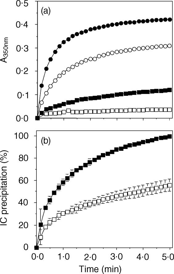
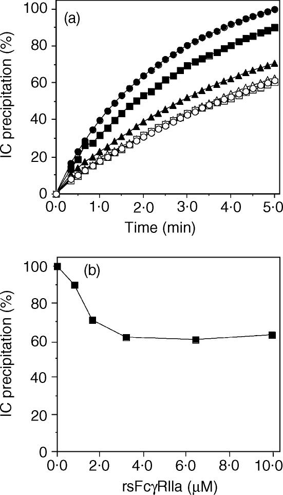
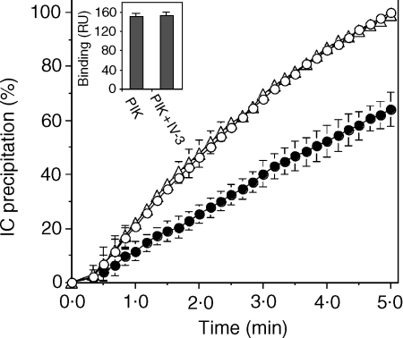
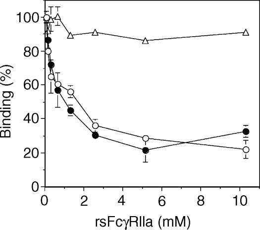
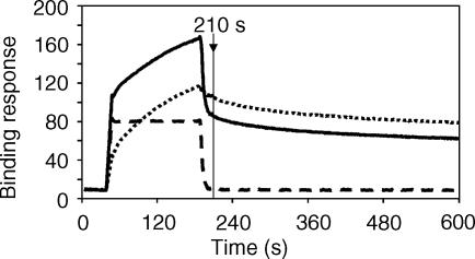
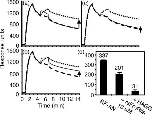
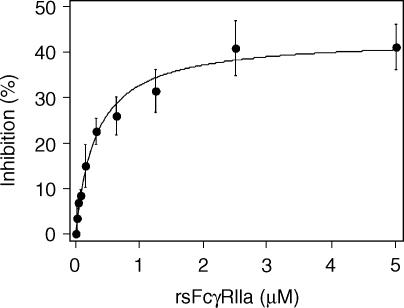
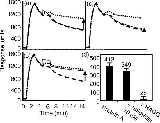
Soluble low-affinity receptors for IgG are known to inhibit immune complex (IC)-mediated inflammation, and expression by leukocytes is elevated in several inflammatory diseases. Immunoglobulin M (IgM) rheumatoid factors (RF), anti-Fc autoantibodies, are found in autoimmune diseases, such as rheumatoid arthritis (RA), as well as in normal immune responses. This study demonstrated that soluble FcgammaRIIa inhibits the interaction of rheumatoid factors with ICs. The recombinant soluble low-affinity FcgammaR, rsFcgammaRIIa, partially inhibited (30-70%) the rate of precipitation of soluble ICs by RF-positive RA sera. This required the normal interaction of FcgammaRIIa with Fc as the effect could be abrogated with the Fab fragment of the blocking mAb IV-3. Furthermore, rsFcgammaRIIa partially inhibited (40%) the binding of a monoclonal IgM RF (RF-AN) to an IC formed by IgG2 antibody binding to an antigen-coated biosensor chip. Since RF-AN has been characterized by crystallography to bind to the CH2/CH3 interface of the IgG-Fc, and leukocyte FcgammaRIIa binds to a distinct site centred on the lower hinge, this inhibition is uncompetitive. Some inhibition (15%) of staphylococcal protein A binding to IC was also observed. As soluble FcgammaRIIa disrupts Fc:Fc interactions in IgG-ICs, we propose that this alteration of the IC also reduces the accessibility of Fc portions in the IC, resulting in the partial inhibition of ligands, particularly IgM RF, which bind Fc. We propose that the high concentrations of soluble FcgammaR found during inflammation can affect the properties of ICs and their interaction with the immune system.


Figures

Figure 1
Figure 1
rsFcγRIIa inhibits RF-mediated precipitation of immune complexes. (a) HAGG (100 µg/ml, 0·7 µm) was incubated with RF-positive serum ELL (squares) or with RF-positive serum TOG (circles) in the absence (filled symbols) or presence (open symbols) of 7·6 µm rsFcγRIIa and complex formation measured by A350nm. (b) Four different RA-positive sera of equivalent RF activity were incubated with HAGG in the absence (filled squares) or presence (open squares) of 7·6 µm rsFcγRIIa as above. The data are presented as the per cent IC precipitation averaged for the four samples ± SD.
Figure 2
Figure 2
rsFcγRIIa inhibition of RF-mediated precipitation is dose dependent. (a) HAGG (100 µg/ml) was incubated with the RF-positive sera (BEA) at 1/40 dilution and complex formation measured in the presence of rsFcγRIIa concentrations of 0 (filled circles), 0·8 (filled squares), 1·7 (filled triangles), 3·3 (open circles), 6·6 (open squares) and 9·9 µm (open triangles). (b) The per cent IC precipitation at the endpoint (5 min) of each assay was plotted against rsFcγRIIa concentration to show that only 40% of the IC precipitation appears sensitive to inhibition by rsFcγRIIa.
Figure 3
Figure 3
The Fab fragment of mAb IV-3 ablates inhibition by rsFcγRIIa of RF immune precipitation. HAGG (100 µg/ml, 0·7 µm) was incubated with a 1/40 dilution of the RF-positive serum PIK alone (open circles), or in the presence of rsFcγRIIa (0·7 µm, closed circles), or in the presence of both rsFcγRIIa and the IV-3 Fab (1·4 µm, open triangles). Values represent the average of four measurements. Insert: PIK serum at 1/40 dilution was bound to HAGG immobilized on a biosensor chip as described in ‘Materials and methods’ and shown in Fig. 5. In the presence of IV-3 mAb (0·6 µm), no effect on RF binding activity was seen (n = 9).
Figure 4
Figure 4
RsFcγRIIa inhibits the binding of HRP-labelled RF to HAGG. HRP-labelled IgM RF BEA (open circles), PIK (closed circles) or protein A (open triangles) was reacted with HAGG-coated ELISA plates as described in the ‘Materials and methods’ in the presence of the indicated concentrations of rsFcγRII. Normalized binding is expressed as a percentage of maximum binding in the absence of rsFcγRIIa. Values represent the average of duplicate measurements.
Figure 5
Figure 5
Biosensor analysis of rsFcγRIIa inhibition of the binding of IgM RF to HAGG. HAGG (∼1000 RU) was coupled to a biosensor chip and the purified IgM (3 µm) from the RA patient PIK was injected (at 210 s, 110 RU bound, sensogram indicated by the dotted line). When the IgM RF preparation was combined with 2 µm rsFcγRIIa in a single injection (solid line), a small but consistent approximate 20% decrease in RF binding was seen at the 210-s time-point. An injection of rsFcγRIIa (2 µm) alone showed that this low-affinity receptor rapidly dissociated from the HAGG layer (dashed line).
Figure 6
Figure 6
RsFcγRIIa inhibits RF-AN binding to immobilized chimeric IgG2. (a, b, c) Chimeric IgG2 anti-NP with human constant domains (12 µg/ml, 80 nm) was injected onto the biosensor (20 µl, 10 µl/min, in 20 mm sodium phosphate, 150 mm NaCl, 2 mm EDTA, 0·05% surfactant p20) over a NP coupled CM5 biosensor chip and binding recorded as response units. An injection of RF-AN (20 µl, 10 µg/ml, 11 nm) was carried out and binding to the IgG2 recorded (dotted line). The chip was regenerated and IgG2 injected as previously, and a second injection of buffer alone carried out to show any non-specific effects and the dissociation of the IgG2 from the chip (dashed line). The binding response of the RF-AN injection above that of the IgG2 injected alone (marked with an arrow) indicates RF-AN binding activity. Following regeneration, other test injections to show binding to the IgG2 layer were carried out and are shown as thin lines. These are the injection of (a) rsFcγRIIa (20 µl, 10 µm), (b) a mixture of rsFcγRIIa (10 µm) and RF-AN (11 nm) and (c) a mixture of heat-aggregated IgG (200 µg/ml, 1·3 µm) and RF-AN (11 nm). Non-specific binding was evaluated by simultaneously passing injected samples over a biosensor channel containing bound chimeric IgE anti-NP. Transient responses on the IgE layer of 11 and 29 RU were recorded during the injection of the RF-AN and rsFcγRIIa samples. (d) Shows the averaged RF binding activity (i.e. the binding response above IgG2 alone) measured at 14 min in the absence of an inhibitor, in the presence of 10 µm rsFcγRIIa and in the presence of 1·3 µm HAGG (average ± SD, n = 4).
Figure 7
Figure 7
RsFcγRIIa inhibition of RF-AN binding activity is concentration dependent. Chimeric IgG2 was reacted with a NP coupled biosensor chip and rsFcγRIIa was used at the indicated concentrations as an inhibitor of RF-AN binding as described for Figure 6 and in the ‘Materials and methods’.
Figure 8
Figure 8
There is little inhibition by rsFcγRIIa of protein A binding to immobilized IgG2. The inhibition of protein A (11 nm) binding to IgG2 by rsFcγRIIa was assessed using the conditions described in Fig. 6. IgG2 was bound to the NP coupled chip (20 µl, 10 ml/min) and ∼4 min later rsProtein A (20 µl, 11 nm) was injected (thick dotted lines) or buffer alone (thick dashed lines) was injected. The binding response of the IgG2 and rsProtein A injection above that of the IgG2 and buffer injection (marked with an arrow) indicates protein A binding activity. Following regeneration, other injections to test binding to the IgG2 layer were carried out and are shown as thin lines. These were the injection of (a) rsFcγRIIa (20 µl, 10 µm), (b) a mixture of rsFcγRIIa (10 µm) and rsProtein A (11 nm), and (c) a mixture of heat-aggregated IgG (200 µg/ml, 1·3 µm) and rsProtein A (11 nm). (d) The average rsProtein A binding activity measured at 14 min in the absence of an inhibitor, in the presence of 10 µm rsFcγRIIa, and in the presence of 1·3 µm HAGG is shown (average ± SD, n = 9).

References

    1. Hulett MD, Hogarth PM. Molecular basis of Fc receptor function. Adv Immunol. 1994;57:1–127. - PubMed
    1. Sylvestre DL, Ravetch JV. Fc receptors initiate the Arthus reaction: redefining the inflammatory cascade. Science. 1994;265:1095–8. - PubMed
    1. Ravetch JV. Fc receptors: rubor redux. Cell. 1994;78:553–60. - PubMed
    1. Ravetch JV, Kinet JP. Fc receptors. Annu Rev Immunol. 1991;9:457–92. - PubMed
    1. Bolland S, Ravetch JV. Spontaneous autoimmune disease in Fc (gamma) RIIB-deficient mice results from strain-specific epistasis. Immunity. 2000;13:277–85. - PubMed

Publication types

MeSH terms