. 2003 Jun;109(2):271-82.
doi: 10.1046/j.1365-2567.2003.01644.x.

Assessment of oral transmission using cell-free human immunodeficiency virus-1 in mice reconstituted with human peripheral blood leucocyte

Affiliations

Assessment of oral transmission using cell-free human immunodeficiency virus-1 in mice reconstituted with human peripheral blood leucocyte

Ryoma Nakao et al. Immunology. 2003 Jun.

Abstract

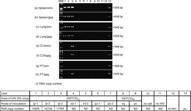
Oral-genital contact is one of the risk factors for the transmission of human immunodeficiency virus (HIV) in adults. In recent reports, oral exposure to simian immunodeficiency virus (SIV) was found to have important implications for the achievement of mucosal transmission of HIV in a rhesus monkey animal model. In the present study, we aimed first to establish a small animal model which did not develop tonsils suitable for HIV oral mucosa transmission, using non-obese diabetic/severe combined immunodeficiency (NOD/SCID) mice and NOD/SCID B2m(null) mice grafted with human peripheral blood leucocytes (hu-PBL) and stimulated with interleukin (IL)-4, and second to investigate whether oral exposure to cell-free R5 and X4 HIV-1 could lead to oral transmission of HIV through intact or traumatized mucosal tissues in humanized mice. Both low and high concentrations of cell-free R5 and X4 viruses failed to cause oral transmission with or without trauma in hu-PBL-NOD/SCID and NOD/SCID Beta2m(null) mice, which presented a number of CD4+ cells in gingival tissues and oral cavities with or without tissue injury. The present results show that IL-4-administrated NOD/SCID B2m(null) mice are useful as a small-humanized model for the study of HIV oral infection, which may help to define the window of opportunity for oral transmission by the HIV virus in animal model experiments.


Figures

Figure 1
Figure 1
Flow cytometric analyses of hu-CD45, CD8, and CD4 T-cell populations present in the spleens of grafted NOD/SCID B2mnull mice treated with hu-IL-4. Hu-PBL in spleen cells were analysed 3 weeks after the last administration using hu-CD45+ single and CD4+/CD8+ double staining. The analyses were performed on gated lymphocytes with forward scatter/side scatter characteristics. Lymphocytes can be distinguished in the quadrant analysis. The number in the upper left and lower right quadrants are the percentages of CD8+ CD4 T cells and CD4+ CD8 T cells, respectively. Determinations of the proportion of non stained cells, hu-CD45+ cells, and hu-CD4+ CD8 and hu-CD4 CD8+ T cells from the spleens of grafted mice given PBS (a) or hu-IL-4 (250 ng/mouse) (b), as well as non-grafted mice (c) are shown. Data shown are representative of 7 (PBS), 14 (IL-4), and 3 (no PBL) independent experiments. No Hu-PBL: spleens from NOD/SCID B2mnull mice.
Figure 2
Figure 2
Effects of hu-IL-4 alone on T-cell proportions in grafted mice infected or not infected with HIV-1. Determinations of the proportion of hu-CD45+ cells, and hu- CD4+ CD8 and hu-CD4 CD8+ T cells from the spleens of grafted mice given PBS and IL-4 (250 ng/mouse) are shown in human PBMC (a), grafted NOD/SCID mice (b), not infected NOD/SCID B2mnull mice (c), and NOD/SCID B2mnull infected with HIV-1 (d). The CD4/CD8 (CD4+ CD8 T cells/CD4 CD8+ T cells) ratio is shown in the right panel. Hu-lymphocytes in spleen cells were analysed 3 weeks after the last administration. Analyses were performed on gated lymphocytes with forward scatter/side scatter characteristics. Results are expressed as means ± SD for representaative independent assays. Statistical analysis was performed by Mann–Whitney's U-test. Asterisks denote significantly different (versus PBS, *P < 0·05, **P < 0·01).
Figure 3
Figure 3
Photomicrographs of pulp, and junction of epithelium and lamina propria specimens from hu-PBL-NOD/SCID B2mnull mice given PBS (× 400). H&E staining of pulp (a), and junction of epithelium and lamina propria (d) are shown in the left column. Immunohistochemical staining of CD45+ cells and non-staining are shown in the pulp (b and c, respectively) and junction of epithelium and lamina propria (e and f, respectively). Data shown are representative of four independent experiments.
Figure 4
Figure 4
Photomicrographs of periodontal tissue specimens from hu-PBL-NOD/SCID mice (× 200). H&E staining (a and b), immunohistochemical staining of hu-CD45+ cells (c and d), and hu-CD4+ (e and f) and hu-CD8+ T cells (g and h) in periodontal tissues from hu-PBL-NOD/SCID mice given PBS and IL-4 are shown. Left and right panels show periodontal sections from mice given PBS and IL-4, respectively, on days 1, 7, and 14. Photos show serial sections in all figures from upper to lower. Data shown are representative of 3 independent experiments.
Figure 5
Figure 5
Photomicrographs of periodontal tissue specimens from hu-PBL-NOD/SCID B2mnull mice (× 200). H-E staining (a and b). Immunohistochemical staining of hu-CD45+ cells (c and d), and hu-CD4+ (e and f) and hu-CD8+ T cells (g and h) in periodontal tissues from hu-PBL-NOD/SCID mice given PBS and IL-4, respectively. Left and right panels show periodontal sections from mice administrated PBS and IL-4, respectively, on days 1, 7, and 14. Photos show serial sections in all figures from upper to lower. Data shown are representative of three independent experiments.
Figure 6
Figure 6
PCR products for env and gag genes in spleen (a and b), lung (c and d), and CLN (e and f) tissues from NOD/SCID mice and NOD/SCID B2mnull mice, respectively, infected with HIV-1. RNA copy numbers are shown in g. Hu-PBL-NOD/SCID mice were inoculated with HIV-1MN via different routes: the peritoneal cavity (lane 1), and oral cavity with (lane 2) and without (lane 3) injured tissues. NOD/SCID B2mnull mice were inoculated with HIV-1MN via different routes: peritoneal cavity (lane 4), and oral cavity with (lanes 5 and 7) and without (lanes 6 and 8) injured tissues. Lane ‘MW’: Molecular weight standards (HaeIIIdigest of ΦX174). As shown in the table (g), the detection limit of RNA copy numbers was 800 in this experiment. Data shown are representative of 3 independent experiments. ND, not detected; ip, intraperitoneal cavity; inj, oral cavity with injured tissues; po, oral cavity; no cell, tissues from NOD/SCID B2mnull mice inoculated intraperitoneally with HIV-1; no HIV, tissues from hu-PBL-NOD/SCID B2mnull mice; no tem, negative control for PCR assay (no template DNA); pc, positive control DNA (HIV-1MN).
Figure 7
Figure 7
PCR products for env and gag genes in spleen (a and b), lung (c and d), CLN (e and f), and PT (g and h) tissues from NOD/SCID mice and NOD/SCID B2mnull mice, respectively, infected with HIV-1. RNA copy numbers are shown in i. Hu-PBL-NOD/SCID mice were inoculated with HIV-1TH22PF7 via different routes: the peritoneal cavity (lanes 1 and 2), and oral cavity with (lane 4) and without (lane 6) tissue injury. Hu-PBL-NOD/SCID B2mnull mice were inoculated with HIV-1JR-FL via different routes: the peritoneal cavity (lane 3) and oral cavity with (lanes 5 and 8) and without (lanes 7 and 9) tissue injury. Lane ‘MW’: Molecular weight standards (HaeIII digest of ΦX174). As shown in the table (i), the detection limit of RNA copy numbers was 800 in this experiment. Data shown are representative of one or two independent experiments. ND, not detected; ip, intraperitoneal cavity; inj, oral cavity with injured tissues; po, oral cavity; no cell: tissues from NOD/SCID B2mnull mice inoculated intraperitoneally with HIV-1; no HIV, tissues from hu-PBL-NOD/SCID B2mnull mice; no tem, negative control for PCR assay (no template DNA); pc, positive control DNA (HIV-1MN).
Figure 8
Figure 8
Flow cytometric analysis of hu-CXCR4+ and CCR5+ cells, and GalCer+ epithelial cell populations in mucosal tissues from the oral cavities of treated NOD/SCID B2mnull mice. The analyses were performed on gated cells larger than lymphocytes with forward scatter/side scatter characteristics. Mucosal tissue cells can be distinguished in the quadrant analysis. The proportions of CXCR4+ and CCR5+ cells were compared to non stained cells as a negative control. The proportion of GalCer+ cells among CD45 cells was compared to cells stained with the control isotype antibody. Data shown are representative of three independent experiments.

Similar articles

Cited by

References

    1. Goedert JJ, Duliege AM, Amos CI, Felton S, Biggar RJ. High risk of HIV-1 infection for first-born twins. The International Registry of HIV-exposed Twins. Lancet. 1991;338:1471–5. - PubMed
    1. Nduati R, John G, Mbori-Ngacha D, et al. Effect of breastfeeding and formula feeding on transmission of HIV-1. JAMA. 2000;283:1167–74. - PubMed
    1. Rothenberg RB, Scarlett M, del Rio C, Reznik D, O'Daniels C. Oral transmission of HIV. AIDS. 1998;12:2095–105. - PubMed
    1. Schacker T, Collier AC, Hughes J, Shea T, Corey L. Clinical and epidemiologic features of primary HIV infection. Ann Intern Med. 1996;125:257–64. - PubMed
    1. Baba TW, Trichel AM, An L, Liska V, Martin LN, Murphey-Corb M, Ruprecht RM. Infection and AIDS in adult macaques after nontraumatic oral exposure to cell-free SIV. Science. 1996;272:1486–9. - PubMed

Publication types

MeSH terms