. 2003 Jun;162(6):1771-80.
doi: 10.1016/S0002-9440(10)64312-X.

Reciprocal modulation of matrix metalloproteinase-13 and type I collagen genes in rat hepatic stellate cells

Affiliations

Reciprocal modulation of matrix metalloproteinase-13 and type I collagen genes in rat hepatic stellate cells

Benjamin Schaefer et al. Am J Pathol. 2003 Jun.

Abstract

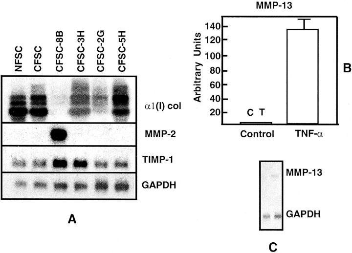
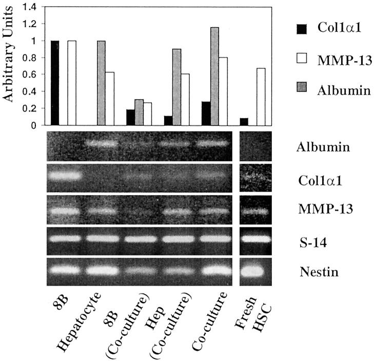
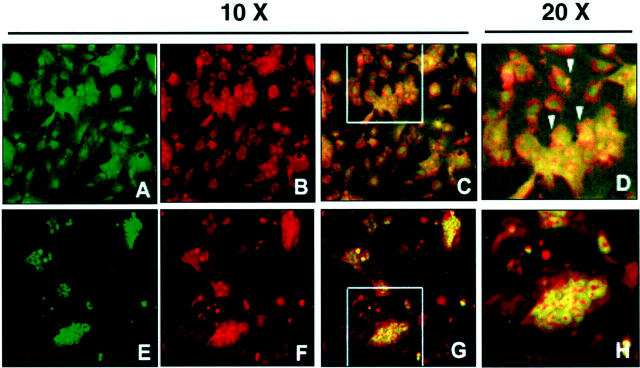
Collagen degradation by matrix metalloproteinases is the limiting step in reversing liver fibrosis. Although collagen production in cirrhotic livers is increased, the expression and/or activity of matrix metalloproteinases could be normal, increased in early fibrosis, or decreased during advanced liver cirrhosis. Hepatic stellate cells are the main producers of collagens and matrix metalloproteinases in the liver. Therefore, we sought to investigate whether they simultaneously produce alpha1(I) collagen and matrix metalloproteinase-13 mRNAs. In this communication we show that expression of matrix metalloproteinase-13 mRNA is reciprocally modulated by tumor necrosis factor-alpha and transforming growth factor-beta1. When hepatic stellate cells are co-cultured with hepatocytes, matrix metalloproteinase-13 mRNA is up-regulated and alpha1(I) collagen is down-regulated. Injuring hepatocytes with galactosamine further increased matrix metalloproteinase-13 mRNA production. Confocal microscopy and differential centrifugation of co-cultured cells revealed that matrix metalloproteinase-13 is localized mainly within hepatic stellate cells. Studies performed with various hepatic stellate cell lines revealed that they are heterogeneous regarding expression of matrix metalloproteinase-13. Those with myofibroblastic phenotypes produce more type I collagen whereas those resembling freshly isolated hepatic stellate cells express matrix metalloproteinase-13. Overall, these findings strongly support the notion that alpha1(I) collagen and matrix metalloproteinase-13 mRNAs are reciprocally modulated.


Figures

Figure 1.
Figure 1.
HSCs are heterogeneous with regard to expression of MMP-2 and MMP-13 mRNAs. Representative Northern blots performed with 10 μg of total RNA extracted from the maternal HSC lines NFSC and CFSC, from four clones derived from CFSC (A), and from CFSC-8B treated with 20 ng/ml of TNF-α (B). The figure shows that while the cell lines and clones expressed variable levels of α1(I) procollagen and TIMP-1 mRNAs, only clone CFSC-8B expressed MMP-2 (A). None of the HSCs tested expressed detectable levels of MMP-9 (data not shown) and only CFSC-8B expressed MMP-13 mRNA whose expression was up-regulated by TNF-α. The histogram in B corresponds to triplicate experiments performed with total RNA extracted 12 hours after culturing cells with TNF-α (see Materials and Methods). Values were corrected for loading differences using GAPDH mRNA as an internal control, and are expressed as means ± SD. C corresponds to a representative Northern blot.
Figure 2.
Figure 2.
PCR analysis of desmin, nestin, glial fibrillary acidic protein, and α-smooth muscle actin in maternal HSC lines (NFSC and CFSC) and in the four clones derived from CFSC (2G, 8B, 3H, and 5H). This figure shows a representative gel of the transcripts detected using total RNA extracted from each of the HSC lines and clones that summarizes the findings obtained. Although the signal intensities varied from cell to cell, all expressed strong signals for most HSC markers. However, levels of expression of glial fibrillary acidic protein were barely detectable in CFSC-8B.
Figure 3.
Figure 3.
Time-dependent expression of MMP-2, TIMP-1, α1(I) procollagen, fibronectin, and CSF-1 mRNAs by cultured CFSC-8B. Northern blot analysis was performed with 10 μg of total RNA. The first time point was obtained with RNA extracted 24 hours after plating HSCs. The zero time point was obtained after trypsinization of confluent cultures before plating. Left: Histograms obtained with data from triplicate experiments. Results are expressed as arbitrary units ± SD and were corrected for loading differences after hybridization with a GAPDH cDNA. Right: A representative Northern blot.
Figure 4.
Figure 4.
Expression of α1(I) procollagen, MMP-2, and MMP-13 mRNAs by co-cultures of CFSC-8B and CFSC-3H. Cells were plated to contain 100%, 75%, 50%, 25%, and 0% CFSC-8B and the expression of the aforementioned mRNAs determined after 12 hours of co-culture. Northern analysis was performed with 10 μg of total RNA as described above. Right: Histograms of triplicate experiments. Results are expressed as fold change compared to controls and are means ± SD after correction for loading differences using GAPDH mRNA as an internal control. Left: A representative blot. Note that MMP-2 mRNA expression reflects the proportion of CFSC-8B in the co-culture, while that of α1(I) procollagen mRNA reflects the number of CFSC-3H. Although TNF-α had no effect on MMP-2 mRNA expression it up-regulated the expression of MMP-13 mRNA.
Figure 5.
Figure 5.
Expression of α1(I) procollagen, MMP-13 (A) and MMP-2 (B) mRNAs by cultured hepatocytes, CFSC-8B, and co-cultures of hepatocytes plus CFSC-8B. Northern blot analysis was performed with 10 μg of total RNA as described in Materials and Methods. Note that while neither hepatocytes nor CFSC-8B cultured alone expressed detectable levels of MMP-13, co-cultures containing both cell types expressed this mRNA and values remained elevated for 24 and 48 hours of co-culture (A). In contrast to these findings, while CFSC-8B expressed high levels of MMP-2 mRNA, this mRNA was not expressed by hepatocytes and its expression level was significantly lower in co-cultures (B). A shows that α1(I) procollagen mRNA expression was down-regulated by 24 and 48 hours of co-culture. Detection of this mRNA was deliberately overexposed to detect the very low levels expressed by co-cultures. Even under these conditions, α1(I) procollagen mRNA was not detected in cultured hepatocytes.
Figure 6.
Figure 6.
Northern (A) and Western blot (B) analyses of MMP-13 expression by co-cultures prepared with various ratios of CFSC-8B cells and hepatocytes. Note that expression of MMP-13 mRNA increased as the ratio of hepatocytes/CFSC-8B was increased in co-culture. Values were maximal above a ratio of 5:1 and remained elevated even at ratios of 10:1. Similarly, the presence of MMP-13 protein in culture media was detectable only with co-cultures containing five hepatocytes/CFSC-8B. Note that while MMP-13 present in the culture media of CFSC-8B cultured alone corresponds to procollagenase, that collected from the co-cultures is the active form. The intensity of the signals do not reflect the actual concentration of MMPs. Western blots correspond to two distinct experiments, different volumes of culture medium were concentrated (10-fold versus 25-fold) and different development times were used to detect the protein.
Figure 7.
Figure 7.
RT-PCR analysis of transcripts of albumin, α1(I) collagen, MMP-13, S14, and nestin expressed by monocultures of CFSC-8B(8B), hepatocytes, freshly isolated HSCs, co-cultures of hepatocytes and CFSC-8B, and CFSC-8B (8B, co-culture), and hepatocytes (Hep Co-culture) isolated after co-culturing the cells for 48 hours. All of the reactions were performed as described using forward and reverse primers of the control gene (S14) and that under investigation. Although signal intensities for the same gene are comparable, signal intensities of the various genes investigated are not. The number of cycles required for optimal amplification of each gene was different (see Materials and Methods). The bars correspond to duplicate experiments. Although the cells isolated after co-culture are contaminated, nonetheless the data show that collagen gene expression by HSCs obtained from co-cultures is very low as compared to monocultured cells. It is noteworthy to indicate that HSCs contaminating the hepatocyte fraction express the highest levels of MMP-13 mRNA.
Figure 8.
Figure 8.
Confocal immunomicroscopy of co-cultures of hepatocytes with CFSC-8B. The cells were co-cultured for 48 hours and fixed as described. The upper row (A–D) corresponds to double-antibody staining for desmin (HSC marker, green) and MMP-13 (red). The lower row (E–H) represents double staining for albumin (hepatocyte marker, green) and MMP-13 (red). A–C and E–G were taken with ×10 magnification. D and H are a ×2 enlargement of the areas indicated in C and G, respectively. Please note that orange-yellow color (arrows) is only observed in HSCs and not in hepatocytes.
Figure 9.
Figure 9.
This figure corresponds to duplicate experiments in which co-cultures of hepatocytes and CFSC-8B were treated with 5 mmol/L of galactosamine. The table shows that treatment with galactosamine induced hepatocyte injury as determined by the increase in LDH activity in the culture medium. The Northern blot is a representative blot showing the increased expression of MMP-13 in galactosamine-treated as compared to control co-cultures.
Figure 10.
Figure 10.
Data presented in this figure support the notion that there is a reciprocal modulation of α1(I) collagen and MMP-13 mRNAs in early passaged (five to six times) mouse HSCs treated with 20 ng/ml of TNF-α or 8 ng/ml of TGF-β1 for 12 hours. The figure illustrates that while TNF-α induced the expression of MMP-13 mRNA it inhibited that of α1(I) collagen, TGF-β1 induced the expression of α1(I) collagen mRNA and down-regulated that of MMP-13.

Similar articles

Cited by

References

    1. Eng FJ, Friedman SL: Fibrogenesis I: new insights into hepatic stellate cell activation: the simple becomes complex. Am J Physiol 2000, 279:G7-G11 - PubMed
    1. Pinzani M, Marra F: Cytokine receptors and signaling in hepatic stellate cells. Semin Liver Dis 2001, 21:397-416 - PubMed
    1. Rockey DC: Hepatic blood flow regulation by stellate cells in normal and injured liver. Semin Liver Dis 2001, 21:337-349 - PubMed
    1. Geerts A: History, heterogeneity, developmental biology, and functions of quiescent hepatic stellate cells. Semin Liver Dis 2001, 21:311-335 - PubMed
    1. Ogata I, Saez CG, Greenwel P, Ponce ML, Geerts A, Leinwand L, Rojkind M: Rat liver fat-storing cell lines express sarcomeric myosin heavy chain mRNA and protein. Cell Motil Cytoskel 1993, 26:125-132 - PubMed

Publication types

MeSH terms

LinkOut - more resources